Preparation And Storage
Recommended Assay Procedures
BD® CompBeads can be used as surrogates to assess fluorescence spillover (Compensation). When fluorochrome conjugated antibodies are bound to BD® CompBeads, they have spectral properties very similar to cells. However, for some fluorochromes there can be small differences in spectral emissions compared to cells, resulting in spillover values that differ when compared to biological controls. It is strongly recommended that when using a reagent for the first time, users compare the spillover on cells and BD® CompBeads to ensure that BD® CompBeads are appropriate for your specific cellular application.
Product Notices
- Since applications vary, each investigator should titrate the reagent to obtain optimal results.
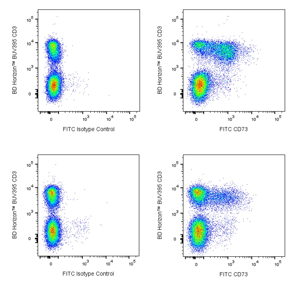
- An isotype control should be used at the same concentration as the antibody of interest.
- Caution: Sodium azide yields highly toxic hydrazoic acid under acidic conditions. Dilute azide compounds in running water before discarding to avoid accumulation of potentially explosive deposits in plumbing.
- Please refer to www.bdbiosciences.com/us/s/resources for technical protocols.
- For fluorochrome spectra and suitable instrument settings, please refer to our Multicolor Flow Cytometry web page at www.bdbiosciences.com/colors.
- Please refer to http://regdocs.bd.com to access safety data sheets (SDS).
Companion Products


The TY/11.8 monoclonal antibody specifically recognizes mouse CD73 which is also known as Ecto-5'-nucleotidase (5'-NT). CD73 is a ~69 kDa glycosylphosphatidylinositol (GPI)-anchored, cell-surface glycoprotein that is encoded by Nt5e which belongs to the 5'-nucleosidase family. CD73 expression appears to be developmentally regulated on leucocytes. In the bone marrow, it is found on most CD11b+ myeloid cells and very few CD19+ cells of the B-lymphocyte lineage. It is neither found expressed on CD11b+ cells in the periphery nor on bone marrow-derived GM-CSF-stimulated dendritic cells. Some peripheral B lymphocytes express CD73, with higher levels detected on Ig isotype-switched B cells. The few thymocytes which have detectable surface CD73 are found within CD4-CD8-and the CD4+CD8- subpopulations. In peripheral lymphoid organs, large proportions of the CD4+ and CD8+ T lymphocytes express CD73. Significant variation in the frequencies of peripheral CD73+ T cells have been observed amongst inbred mouse strains. For example, C57BL/6 mice reportedly have higher frequencies of peripheral CD73+ T cells when compared with BALB/c mice. In the thymus and peripheral lymphoid organs, CD73 is found on endothelial and stromal cells. CD73 has also been detected on bone marrow and thymic epithelial cell lines, kidney glomeruli and proximal-tubule epithelial cells, liver endothelial cells and hepatocytes, mesenchymal cells, and fibroblasts including cancer-associated fibroblasts (CAFs) found in tumors. CD73 has enzymatic and signal transduction activities. It catalyzes the dephosphorylation of extracellular nucleoside 5' monophosphates to nucleosides. CD73 acts on adenosine monophosphate (AMP) to generate and regulate the concentration of extracellular adenosine. Adenosine can bind to adenosine receptors expressed on cells in many tissues and regulate physiological responses including anti-inflammatory or immunosuppressive responses. Regulatory T cells (Treg) can generate immunosuppressive adenosine by their expression and activity of the CD39 and CD73 ectoenzymes.

Development References (6)
-
Barron L, Dooms H, Hoyer KK, et al. Cutting edge: mechanisms of IL-2-dependent maintenance of functional regulatory T cells.. J Immunol. 2010; 185(11):6426-30. (Clone-specific: Flow cytometry). View Reference
-
Deaglio S, Dwyer KM, Gao W, et al. Adenosine generation catalyzed by CD39 and CD73 expressed on regulatory T cells mediates immune suppression.. J Exp Med. 2007; 204(6):1257-65. (Biology). View Reference
-
Le Texier L, Lineburg KE, Cao B, et al. Autophagy-dependent regulatory T cells are critical for the control of graft-versus-host disease. JCI Insight. 2016; 1(15):e86850. (Clone-specific: Flow cytometry). View Reference
-
Resta R, Yamashita Y, Thompson LF. Ecto-enzyme and signaling functions of lymphocyte CD73. Immunol Rev. 1998; 161:95-109. (Biology). View Reference
-
Yamashita Y, Hooker SW, Jiang H, et al. CD73 expression and fyn-dependent signaling on murine lymphocytes. Eur J Immunol. 1998; 28(10):2981-2990. (Immunogen: Flow cytometry). View Reference
-
Yu M, Guo G, Huang L, et al. CD73 on cancer-associated fibroblasts enhanced by the A(2B)-mediated feedforward circuit enforces an immune checkpoint. Nature Commun. 2020; 11(1):515. (Clone-specific: Flow cytometry). View Reference
Please refer to Support Documents for Quality Certificates
Global - Refer to manufacturer's instructions for use and related User Manuals and Technical data sheets before using this products as described
Comparisons, where applicable, are made against older BD Technology, manual methods or are general performance claims. Comparisons are not made against non-BD technologies, unless otherwise noted.
For Research Use Only. Not for use in diagnostic or therapeutic procedures.
Refer to manufacturer's instructions for use and related User Manuals and Technical Data Sheets before using this product as described.
Comparisons, where applicable, are made against older BD technology, manual methods or are general performance claims. Comparisons are not made against non-BD technologies, unless otherwise noted.