
Preparation And Storage
Product Notices
- This reagent has been pre-diluted for use at the recommended Volume per Test. We typically use 1 × 10^6 cells in a 100-µl experimental sample (a test).
- An isotype control should be used at the same concentration as the antibody of interest.
- Caution: Sodium azide yields highly toxic hydrazoic acid under acidic conditions. Dilute azide compounds in running water before discarding to avoid accumulation of potentially explosive deposits in plumbing.
- Source of all serum proteins is from USDA inspected abattoirs located in the United States.
- Pacific Blue™ is a trademark of Molecular Probes, Inc., Eugene, OR.
- For fluorochrome spectra and suitable instrument settings, please refer to our Multicolor Flow Cytometry web page at www.bdbiosciences.com/colors.
- Please refer to www.bdbiosciences.com/us/s/resources for technical protocols.
Companion Products


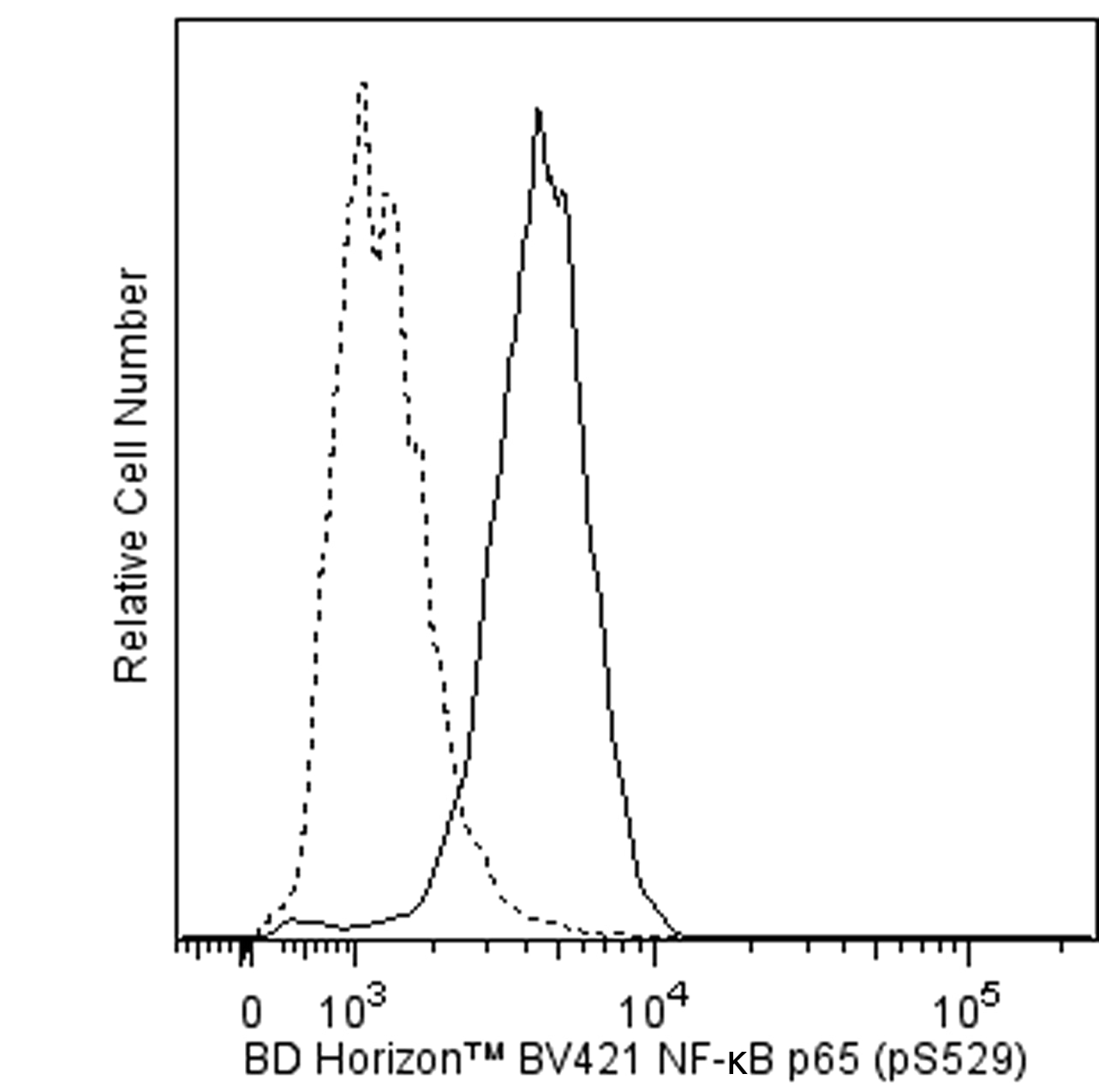
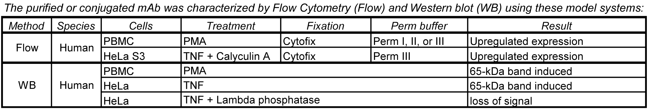
The K10-895.12.50 monoclonal antibody recognizes the phosphorylated serine 529 (pS529) in the transactivation domain of the human NF-κB p65 subunit. Nuclear factor κB (NF-κB) is a ubiquitously expressed transcription factor that regulates the expression of many other genes. It is crucial for cellular responses to a variety of stimuli including stress and microbial pathogens that lead to immunity, inflammation, proliferation, differentiation, survival, apoptosis, and tumorigenesis. The most studied NF-κB complex consists of the p50 (also known as NF-κB1) and p65 (also known as REL-A) subunits, both containing a 300-amino acid region with homology to the Rel proto-oncogene product (RH domain). The RH domain contains motifs for dimerization, nuclear localization, and binding to specific DNA sequences. In addition to the RH domain, the p65 subunit contains the transactivation domain, which is responsible for the interaction with the inhibitor IκB and which contains phosphorylation sites. In most cell types, the p50/p65 heterodimer is located within the cytoplasm complexed to IκB. This complex prevents nuclear translocation and activity of NF-κB. In response to stimuli such as cytokines, LPS, DNA damage, and microbial infections, IκB is phosphorylated at critical residues. This phosphorylation induces dissociation of the IκB/NF-κB complex, allowing the free heterodimeric NF-κB to translocate to the nucleus. Furthermore, optimal activation of NF-κB requires phosphorylation in the transactivation domain of p65. In the nucleus, activated NF-κB dimers bind to the κB sites within promoters and enhancers and function as transcriptional regulators.
The antibody was conjugated to BD Horizon BV421 which is part of the BD Horizon Brilliant™ Violet family of dyes. With an Ex Max of 407-nm and Em Max at 421-nm, BD Horizon BV421 can be excited by the violet laser and detected in the standard Pacific Blue™ filter set (eg, 450/50-nm filter). BD Horizon BV421 conjugates are very bright, often exhibiting a 10 fold improvement in brightness compared to Pacific Blue conjugates.

Development References (8)
-
Dominguez-Villar M, Gautron AS, de Marcken M, Keller MJ, Hafler DA. TLR7 induces anergy in human CD4+ T cells. Nat Immunol. 2015; 16(1):118-128. (Clone-specific: Flow cytometry). View Reference
-
Feasibility study: phospho-specific flow cytometry enabling rapid functional analysis of bone marrow samples from patients with multiple myeloma. Cytometry B Clin Cytom. 2014; 86B:139-144. (Clone-specific: Flow cytometry). View Reference
-
Mingueneau M, Kreslavsky T, Gray D, . The transcriptional landscape of alphabeta T cell differentiation. Nat Immunol. 2013; 14(6):619-632. (Clone-specific: Flow cytometry). View Reference
-
Natoli G, Saccani S, Bosisio D, Marazzi I. Interactions of NF-kappaB with chromatin: the art of being at the right place at the right time. Nat Immunol. 2005; 6(5):439-445. (Biology). View Reference
-
Siebenlist U, Brown K, Claudio E. Control of lymphocyte development by nuclear factor-kappaB. Nat Rev Immunol. 2005; 5:435-445. (Biology). View Reference
-
Suni MA, Maino VC. Flow cytometric analysis of cell signaling proteins. Methods Mol Biol. 2011; 717:155-169. (Clone-specific: Flow cytometry). View Reference
-
Viatour P, Merville M-P, Bours V, Chariot A. Phosphorylation of NF-kappaB and IkappaB proteins: implications in cancer and inflammation. Trends Biochem Sci. 2005; 30(1):43-52. (Biology). View Reference
-
van de Laar L, van den Bosch A, Boonstra A, et al. PI3K-PKB hyperactivation augments human plasmacytoid dendritic cell development and function. Blood. 2012; 120(25):4982-4991. (Clone-specific: Flow cytometry). View Reference
Please refer to Support Documents for Quality Certificates
Global - Refer to manufacturer's instructions for use and related User Manuals and Technical data sheets before using this products as described
Comparisons, where applicable, are made against older BD Technology, manual methods or are general performance claims. Comparisons are not made against non-BD technologies, unless otherwise noted.
For Research Use Only. Not for use in diagnostic or therapeutic procedures.
Refer to manufacturer's instructions for use and related User Manuals and Technical Data Sheets before using this product as described.
Comparisons, where applicable, are made against older BD technology, manual methods or are general performance claims. Comparisons are not made against non-BD technologies, unless otherwise noted.