BD Pharmingen™ Mouse Th1/Th17 Phenotyping Kit
(RUO)

Description
Components:
51-9006632 Mouse Th1/Th17 Phenotyping Cocktail 1.0 ml
Containing the following:
Mouse CD4 PerCP-Cy5.5 (clone: RM4-5)
Mouse IL-17A PE (clone: TC11-18H10.1)
Mouse IFN-GMA FITC (clone: XMG1.2)
51-9006613 BD Cytofix™ Fixation Buffer 100 ml
51-2091KE BD Perm/Wash™ Buffer 25 ml
51-2092KZ BD GolgiStop™ Protein Transport Inhibitor (containing monensin) 0.7 ml
The peripheral CD4+ T cell pool includes multiple effector and memory T cell subsets that arise through antigen-driven expansion and differentiation of naïve T cells. The early response of naïve CD4+ T cells to antigenic stimulation is characterized by high level proliferation and a limited cytokine repertoire. Further differentiation yields cells with a more diverse potential for cytokine expression. Depending upon the balance of local cytokines, costimulatory molecules, antigen levels, and genetic factors, Type-1 T helper (Th1), Th2, and Th17 effector and/or memory cells are generated by immune responses.
Functionally-polarized CD4+ T cell subsets have been identified based on their distinctive patterns of cytokine secretion. As a signature cytokine, Th1 cells selectively produce large amounts of interferon-gamma (IFN-γ). Th2 cells selectively produce IL-4, and Th17 express high levels of IL-17A. Through secretion of IFN-γ and other effector molecules, Th1 cells activate macrophages, natural killer (NK) cells, and CD8+ T cells and are responsible for cell-mediated immunity. Th1 cells provide protection against intracellular bacteria, fungi, protozoa and viruses and are involved in some autoimmune responses. IL-4 produced by Th2 cells is particularly strong in driving B cells to generate IgE-secreting cells. IgE plays a role in basophil/mast cell mediated immune reactions. Th2 cells mediate protection against extracellular parasites but may also cause harmful allergic responsiveness to develop. Through the secretion of IL-17A and other factors, Th17 cells recruit and activate neutrophils and mediate immune responses against extracellular bacteria and fungi. Th17 cells are also implicated in autoimmune responses. In addition to these types of T helper cells, Th0- (IL-4 and IFN-γ) and Th17/Th1-like (IL-17A/IFN-γ) cells that coexpress signature cytokines have been described.
The Th1/Th17 paradigm provides a useful model system for investigating the cellular and molecular mechanisms that mediate protective as well as harmful immune responses. The BD Mouse Th1/Th17 Phenotyping Kit provides an easy-to-use four-color cocktail of fluorescent antibodies-specific for mouse CD4, IFN-γ (for Th1), IL-4 (for Th2) and IL-17A (for Th17)-that will enable researchers to identify and characterize the nature of these T helper cell types by multicolor flow cytometric analysis. The kit can be used to successfully analyze ex vivo lymphoid cell samples (eg, for the types of in vivo-generated T helper cells) or to monitor T helper cell differentiation by cells cultured within various experimental model systems.
Investigators should note that the appearance of BD GolgiStop™ Protein Transport Inhibitor may range in color from clear (colorless) to light yellow.
Preparation And Storage
Recommended Assay Procedures
A. Stimulation of the Cells
Various in vitro methods have been reported for polarization or stimulation of T helper cell subsets of which PMA (Phorbol ester) plus Ionomycin (Calcium Ionophore) has been particularly useful for quickly inducing and characterizing polyclonal cytokine-producing cells. For this kit we recommend the stimulation of mouse spleen cells at a concentration of 1-10 million cells per ml in media for 5 hours with PMA/Ionomycin (at 50 ng/ml and 1 μg/ml respectively) in the presence of BD GolgiStop™ Protein Transport Inhibitor (provided in the kit or Cat #554724). Add 4 μl of BD GolgiStop™ for every 6 ml of cell culture and mix thoroughly. It is recommended that BD GolgiStop™ not be kept in cell culture for longer than 12 hours.
Note: Kinetic studies need to be performed to determine the optimal incubation time for each experimental system. Depending on the mouse, frequencies of cytokine producing cells derived from activation of mouse spleen cells can vary widely for a specific cytokine. In particular, the number of IL-17 producing cells can be very low or even negligible on PMA/Ionomycin stimulated spleen cell. In these cases, Th17 polarization cultures should be considered. For specifics on polarization of Th17 cells please refer to the references.
B. Staining of the Cells
1. Harvest of the Cells
Collect cells from in vitro stimulatory cultures treated with a protein transport inhibitor. Spin down cells at 250 x g for 10 minutes at room temperature (RT) and wash two times with stain buffer (FBS) (Cat# 554656). Count cells and transfer approximately 1 million cells to each flow test tube (Cat# 352008) for immunofluorescent staining. Cells should be protected from light throughout the staining procedure and storage.
2. Fixing the Cells
a. Spin down cells at 250 x g for 10 minutes at RT and thoroughly suspend cells with 1ml of cold BD Cytofix™ buffer (provided in the kit or Cat# 554655) and incubate for 10-20 minutes at RT.
Note: Cell aggregation can be avoided by vortexing prior to the addition of the fixative.
b. Spin down cells at 250 x g for 10 minutes at RT.
Note: After fixation, the cell pellet after centrifugation is loose and care should be taken when aspirating the wash buffer from the tubes. Do not aspirate ALL of the buffer but leave 50-150 µl of solution in the tubes to avoid cell loss for all subsequent wash steps below.
c. Wash cells twice at RT in stain buffer (FBS) and spin down the cells at 250 x g for 10 minutes at RT.
Note: Cells can be stored in stain buffer at 4°C for up to 72 hours or in 90% FCS/10% DMSO at -80°C for up to six months (for samples that need to be stored longer than six months, we recommend performing stability studies) .
3. Permeabilizing the Fixed Cells
a. For cells kept at 4°C, spin down cells at 250 x g for 10 minutes at RT and remove stain buffer.
b. For cells stored at -80°C thaw and wash twice with stain buffer (FBS) to remove DMSO.
c. Dilute 10x BD Perm/Wash™ buffer (provided in the kit or Cat# 554723) in distilled water to make a 1x solution prior to use.
d. Suspend cells in 1ml of 1x BD Perm/Wash™ buffer and incubate at RT for 15 minutes.
e. Spin down cells at 250 x g for 10 minutes at RT and remove supernatant.
4. Staining with the Cocktail
a. Thoroughly suspend fixed/permeabilized cells in each tube in 50 μl of BD Perm/Wash™ buffer and add 20 μl/tube of cocktail or appropriate negative control. Incubate at RT for 30 minutes in the dark. Cells should be protected from light throughout the staining procedure.
b. Optional: Staining of Additional Cell Surface Antigens
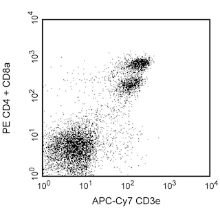
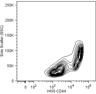
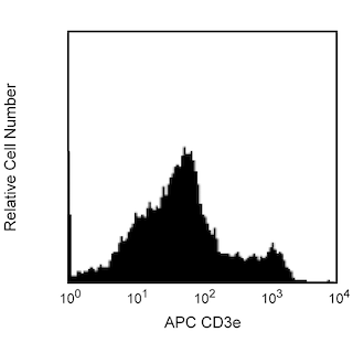
Note: If instrument allows, optional multicolor staining of different cell surface antigens can be done at this time. Example: APC-Cy™7 CD3 (Clone 145-2C11, Cat# 557596) and V450 CD44 (CloneIM7, Cat# 560451) etc. For cell surface staining after fixation/permeabilization, suitable antibody clones that recognize denatured epitopes need to be identified.
Note: For antibodies that do not recognize fixed/denatured cell surface markers, it is recommended that staining be done on live cells PRIOR to fixation/permeabilization.
c. Wash cells twice with 1ml of 1× BD Perm/Wash™ buffer at RT and suspend in Stain Buffer (FBS) prior to flow-cytometric analysis.
C. Flow Cytometric Analysis
Set PMT voltage and compensation using unstained cells and appropriate cell surface markers or use BD™ Compensation beads (Cat #552844) as per the recommended protocol.
Note: It has been reported that CD4 expression on T cells is decreased after cell activation.
Note: Acquire at least 20,000 to 30,000 CD4 positive lymphocytes. Depending on the donor, frequencies of cytokine producing cells derived from activation of human PBMCs can vary widely for a particular cytokine. In order to make statistically significant frequency measurements, sufficiently large sample sizes should be acquired during flow cytometric analysis. Bivariate dot plots or probability contour plots can be generated upon data reanalysis to display the frequencies of and patterns by which individual cells co-express certain levels of cell surface antigen and intracellular cytokine proteins.
Warnings & Precautions
Danger: BD GolgiStop™ Protein Transport Inhibitor (component 51-2092KZ) contains 99.61% ethanol (w/w) and 0.26% monensin, mononatriumsalz (w/w).
Hazard statements:
Highly flammable liquid and vapor.
Causes serious eye irritation.
Precautionary statements:
Keep away from heat/sparks/open flames/hot surfaces. No smoking.
Wear protective gloves / eye protection.
Wear protective clothing.
IF ON SKIN (or hair): Remove / Take off immediately all contaminated clothing. Rinse skin with water / shower.
IF IN EYES: Rinse cautiously with water for several minutes. Remove contact lenses, if present and easy to do. Continue rinsing.
Dispose of contents / container in accordance with local / regional / national / international regulations.
Danger: BD Cytofix™ Fixation Buffer (component 51-9006613) contains 4.2% (w/w) formaldehyde.
Hazard statements:
Harmful if inhaled.
Causes skin irritation.
Causes serious eye damage.
May cause an allergic skin reaction.
Suspected of causing genetic defects.
May cause cancer. Route of exposure: Inhalative.
May cause respiratory irritation.
Precautionary statements:
Wear protective clothing / eye protection.
Wear protective gloves.
Do not breathe mist/vapours/spray.
IF IN EYES: Rinse cautiously with water for several minutes. Remove contact lenses, if present and easy to do.
Continue rinsing.
If skin irritation or rash occurs: Get medical advice/attention.
Product Notices
- Please observe the following precautions: Absorption of visible light can significantly alter the energy transfer occurring in any tandem fluorochrome conjugate; therefore, we recommend that special precautions be taken (such as wrapping vials, tubes, or racks in aluminum foil) to prevent exposure of conjugated reagents, including cells stained with those reagents, to room illumination.
- PerCP-Cy5.5 is optimized for use with a single argon ion laser emitting 488-nm light. Because of the broad absorption spectrum of the tandem fluorochrome, extra care must be taken when using dual-laser cytometers, which may directly excite both PerCP and Cy5.5™. We recommend the use of cross-beam compensation during data acquisition or software compensation during data analysis.
- PerCP-Cy5.5–labelled antibodies can be used with FITC- and R-PE–labelled reagents in single-laser flow cytometers with no significant spectral overlap of PerCP-Cy5.5, FITC, and R-PE fluorescence.
- This PerCP-conjugated product is sold under license to the following patent: US Patent No. 4,876,190.
- Cy is a trademark of Amersham Biosciences Limited. This conjugated product is sold under license to the following patents: US Patent Nos. 5,486,616; 5,569,587; 5,569,766; 5,627,027.
- This product is subject to proprietary rights of Amersham Biosciences Corp. and Carnegie Mellon University and made and sold under license from Amersham Biosciences Corp. This product is licensed for sale only for research. It is not licensed for any other use. If you require a commercial license to use this product and do not have one return this material, unopened to BD Biosciences, 10975 Torreyana Rd, San Diego, CA 92121 and any money paid for the material will be refunded.
- Source of all serum proteins is from USDA inspected abattoirs located in the United States.
- Caution: Sodium azide yields highly toxic hydrazoic acid under acidic conditions. Dilute azide compounds in running water before discarding to avoid accumulation of potentially explosive deposits in plumbing.
- For fluorochrome spectra and suitable instrument settings, please refer to our Multicolor Flow Cytometry web page at www.bdbiosciences.com/colors.
- Please refer to www.bdbiosciences.com/us/s/resources for technical protocols.
Companion Products


.png?imwidth=320)
Development References (6)
-
Carlson MJ, West ML, Coghill JM, et al. In vitro-differentiated TH17 cells mediate lethal acute graft-versus-host disease with severe cutaneous and pulmonary pathologic manifestations. Blood. 2009; 113:1365-1374. (Biology). View Reference
-
Dong C. Th17 cells: Current understanding of their generation and regulation. Eur J Immunol. 2009; 39(3):640-644. (Biology). View Reference
-
Liang SC, Tan XY, Luxenberg DP, et al. Interleukin (IL)-22 and IL-17 are coexpressed by Th17 cells and cooperatively enhance expression of antimicrobial peptides. J Exp Med. 2006; 203:2271-2279. (Biology). View Reference
-
Mosmann TR, Coffman RL. TH1 and TH2 cells: different patterns of lymphokine secretion lead to different functional properties. Annu Rev Immunol. 1989; 7:145-173. (Biology). View Reference
-
Zheng Y, Danilenko DM, Valdez P, et al. Interleukin-22, a T(H)17 cytokine, mediates IL-23-induced dermal inflammation and acanthosis. Nature. 2007; 445:648-651. (Biology). View Reference
-
Zhu J, Paul WE. CD4 T cells: fates, functions, and faults. Blood. 2008; 112(5):1557-1569. (Biology). View Reference
Please refer to Support Documents for Quality Certificates
Global - Refer to manufacturer's instructions for use and related User Manuals and Technical data sheets before using this products as described
Comparisons, where applicable, are made against older BD Technology, manual methods or are general performance claims. Comparisons are not made against non-BD technologies, unless otherwise noted.
For Research Use Only. Not for use in diagnostic or therapeutic procedures.
Refer to manufacturer's instructions for use and related User Manuals and Technical Data Sheets before using this product as described.
Comparisons, where applicable, are made against older BD technology, manual methods or are general performance claims. Comparisons are not made against non-BD technologies, unless otherwise noted.