Old Browser
This page has been recently translated and is available in French now.
Looks like you're visiting us from {countryName}.
Would you like to stay on the current country site or be switched to your country?


Regulatory Status Legend
Any use of products other than the permitted use without the express written authorization of Becton, Dickinson and Company is strictly prohibited.
Preparation And Storage
Recommended Assay Procedures
BD™ CompBeads can be used as surrogates to assess fluorescence spillover (Compensation). When fluorochrome conjugated antibodies are bound to BD CompBeads, they have spectral properties very similar to cells. However, for some fluorochromes there can be small differences in spectral emissions compared to cells, resulting in spillover values that differ when compared to biological controls. It is strongly recommended that when using a reagent for the first time, users compare the spillover on cells and BD CompBead to ensure that BD CompBeads are appropriate for your specific cellular application.
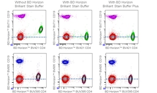
For optimal and reproducible results, BD Horizon Brilliant Stain Buffer should be used anytime two or more BD Horizon Brilliant dyes are used in the same experiment. Fluorescent dye interactions may cause staining artifacts which may affect data interpretation. The BD Horizon Brilliant Stain Buffer was designed to minimize these interactions. More information can be found in the Technical Data Sheet of the BD Horizon Brilliant Stain Buffer (Cat. No. 563794/566349) or the BD Horizon Brilliant Stain Buffer Plus (Cat. No. 566385).
Note: When using high concentrations of antibody, background binding of this dye to erythroid cell subsets (mature erythrocytes and precursors) has been observed. For researchers studying these cell populations, or in cases where light scatter gating does not adequately exclude these cells from the analysis, this background may be an important factor to consider when selecting reagents for panel(s).
Product Notices
- The production process underwent stringent testing and validation to assure that it generates a high-quality conjugate with consistent performance and specific binding activity. However, verification testing has not been performed on all conjugate lots.
- Researchers should determine the optimal concentration of this reagent for their individual applications.
- An isotype control should be used at the same concentration as the antibody of interest.
- Caution: Sodium azide yields highly toxic hydrazoic acid under acidic conditions. Dilute azide compounds in running water before discarding to avoid accumulation of potentially explosive deposits in plumbing.
- For fluorochrome spectra and suitable instrument settings, please refer to our Multicolor Flow Cytometry web page at www.bdbiosciences.com/colors.
- Please refer to www.bdbiosciences.com/us/s/resources for technical protocols.
- BD Horizon Brilliant Stain Buffer is covered by one or more of the following US patents: 8,110,673; 8,158,444; 8,575,303; 8,354,239.
- Please refer to http://regdocs.bd.com to access safety data sheets (SDS).
- CF™ is a trademark of Biotium, Inc.
- BD Horizon Brilliant Ultraviolet 563 is covered by one or more of the following US patents: 8,110,673; 8,158,444; 8,227,187; 8,575,303; 8,354,239.
Companion Products


The R3-242 monoclonal antibody specifically binds to aminopeptidase N (APN), which has been reported to be the mouse CD13 homologue. APN is expressed in a variety of mammalian tissues, including intestinal and renal epithelial brush border, endothelium, liver, lung, and brain. In lymphoid tissues, R3-242 mAb is reported to detect APN on monocytes, macrophages, mast cells, dendritic cells, and B lymphocytes, but not on T cells, and thymocytes. However, the reported reactivity with B cells may be non-specific, via binding to the Fcγ receptors (CD16/CD32). This observation is in agreement with reports using different antibodies to mouse APN. APN may play a role in antigen presentation in the immune system. R3-242 mAb does not inhibit the enzymatic activity of APN.
The antibody was conjugated to BD Horizon™ BUV563 which is part of the BD Horizon Brilliant™ Ultraviolet family of dyes. This dye is a tandem fluorochrome of BD Horizon BUV395 which has an Ex Max of 348 nm and an acceptor dye. The tandem has an Em Max at 563 nm. BD Horizon BUV563 can be excited by the 355 nm ultraviolet laser. On instruments with a 561 nm Yellow-Green laser, the recommended bandpass filter is 585/15 nm with a 535 nm long pass to minimize laser light leakage. When BD Horizon BUV563 is used with an instrument that does not have a 561 nm laser, a 560/40 nm filter with a 535 nm long pass may be more optimal. Due to the excitation and emission characteristics of the acceptor dye, there may be spillover into the PE and PE-CF594 detectors. However, the spillover can be corrected through compensation as with any other dye combination.

Development References (4)
-
Chen H, Kinzer CA, Paul WE. p161, a murine membrane protein expressed on mast cells and some macrophages, is mouse CD13/aminopeptidase N. J Immunol. 1996; 157(6):2593-2600. (Clone-specific: Flow cytometry). View Reference
-
Hansen AS, Noren O, Sjostrom H, Werdelin O. A mouse aminopeptidase N is a marker for antigen-presenting cells and appears to be co-expressed with major histocompatibility complex class II molecules. Eur J Immunol. 1993; 23(9):2358-2364. (Immunogen: Blocking, ELISA, Flow cytometry, Functional assay, Immunoprecipitation, Radioimmunoassay). View Reference
-
Leenen PJ, Melis M, Kraal G, Hoogeveen AT, Van Ewijk W. The monoclonal antibody ER-BMDM1 recognizes a macrophage and dendritic cell differentiation antigen with aminopeptidase activity. Eur J Immunol. 1992; 22(6):1567-1572. (Biology). View Reference
-
Pulendran B, Lingappa J, Kennedy MK, et al. Developmental pathways of dendritic cells in vivo: distinct function, phenotype, and localization of dendritic cell subsets in FLT3 ligand-treated mice. J Immunol. 1997; 159(5):2222-2231. (Biology). View Reference
Please refer to Support Documents for Quality Certificates
Global - Refer to manufacturer's instructions for use and related User Manuals and Technical data sheets before using this products as described
Comparisons, where applicable, are made against older BD Technology, manual methods or are general performance claims. Comparisons are not made against non-BD technologies, unless otherwise noted.
For Research Use Only. Not for use in diagnostic or therapeutic procedures.
Report a Site Issue
This form is intended to help us improve our website experience. For other support, please visit our Contact Us page.