-
Your selected country is
Italy
- Change country/language
Old Browser
This page has been recently translated and is available in French now.
Looks like you're visiting us from {countryName}.
Would you like to stay on the current country site or be switched to your country?
Regulatory Status Legend
Any use of products other than the permitted use without the express written authorization of Becton, Dickinson and Company is strictly prohibited.
Preparation And Storage
Recommended Assay Procedures
For optimal and reproducible results, BD Horizon Brilliant Stain Buffer should be used anytime two or more BD Horizon Brilliant dyes (including BD OptiBuild Brilliant reagents) are used in the same experiment. Fluorescent dye interactions may cause staining artifacts which may affect data interpretation. The BD Horizon Brilliant Stain Buffer was designed to minimize these interactions. More information can be found in the Technical Data Sheet of the BD Horizon Brilliant Stain Buffer (Cat. No. 563794).
Product Notices
- Researchers should determine the optimal concentration of this reagent for their individual applications.
- This antibody was developed for use in flow cytometry.
- An isotype control should be used at the same concentration as the antibody of interest.
- The production process underwent stringent testing and validation to assure that it generates a high-quality conjugate with consistent performance and specific binding activity. However, verification testing has not been performed on all conjugate lots.
- Caution: Sodium azide yields highly toxic hydrazoic acid under acidic conditions. Dilute azide compounds in running water before discarding to avoid accumulation of potentially explosive deposits in plumbing.
- Cy is a trademark of Amersham Biosciences Limited.
- Alexa Fluor® is a registered trademark of Life Technologies Corporation.
- BD Horizon Brilliant Stain Buffer is covered by one or more of the following US patents: 8,110,673; 8,158,444; 8,575,303; 8,354,239.
- BD Horizon Brilliant Violet 711 is covered by one or more of the following US patents: 8,110,673; 8,158,444; 8,227,187; 8,455,613; 8,575,303; 8,354,239.
- Please refer to www.bdbiosciences.com/us/s/resources for technical protocols.
- For fluorochrome spectra and suitable instrument settings, please refer to our Multicolor Flow Cytometry web page at www.bdbiosciences.com/colors.
Companion Products
The GoH3 monoclonal antibody specifically binds to CD49f which is also known as integrin α6 chain. CD49f is a ~150 kDa type I transmembrane glycoprotein that belongs to the integrin alpha chain family of extracellular matrix and cell adhesion receptors. The integrin α6 subunit associates with the integrin β1 chain (CD29) to form VLA-6 and with the integrin β4 chain (CD104) to form the integrin α6β4 complex, also known as the laminin and kalinin receptor. CD49f is expressed mainly on T cells, monocytes, platelets, epithelial cells, endothelial cells, perineural cells, and trophoblasts of placenta. GoH3 recognizes an extracellular epitope of integrin α6 on human, mouse and bovine cells. GoH3 has been reported to block the binding of integrin α6 to laminin P1 and E8 fragments.
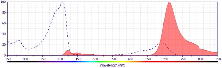
The antibody was conjugated to BD Horizon™ BV711 which is part of the BD Horizon Brilliant™ Violet family of dyes. This dye is a tandem fluorochrome of BD Horizon BV421 with an Ex Max of 405-nm and an acceptor dye with an Em Max at 711-nm. BD Horizon BV711 can be excited by the violet laser and detected in a filter used to detect Cy™5.5 / Alexa Fluor® 700-like dyes (eg, 712/20-nm filter). Due to the excitation and emission characteristics of the acceptor dye, there may be moderate spillover into the Alexa Fluor® 700 and PerCP-Cy5.5 detectors. However, the spillover can be corrected through compensation as with any other dye combination.
Development References (3)
-
Aumailley M, Timpl R, Sonnenberg A. Antibody to integrin alpha 6 subunit specifically inhibits cell-binding to laminin fragment 8. Exp Cell Res. 1990; 188(1):55-60. (Biology). View Reference
-
Knapp W. W. Knapp .. et al., ed. Leucocyte typing IV : white cell differentiation antigens. Oxford New York: Oxford University Press; 1989:1-1182.
-
Sonnenberg A, Daams H, Van der Valk MA, Hilkens J, Hilgers J. Development of mouse mammary gland: identification of stages in differentiation of luminal and myoepithelial cells using monoclonal antibodies and polyvalent antiserum against keratin. J Histochem Cytochem. 1986; 34(8):1037-1046. (Immunogen: Immunohistochemistry, Radioimmunoassay). View Reference
Please refer to Support Documents for Quality Certificates
Global - Refer to manufacturer's instructions for use and related User Manuals and Technical data sheets before using this products as described
Comparisons, where applicable, are made against older BD Technology, manual methods or are general performance claims. Comparisons are not made against non-BD technologies, unless otherwise noted.
For Research Use Only. Not for use in diagnostic or therapeutic procedures.
Report a Site Issue
This form is intended to help us improve our website experience. For other support, please visit our Contact Us page.