Preparation And Storage
Recommended Assay Procedures
For optimal and reproducible results, BD Horizon Brilliant Stain Buffer should be used anytime two or more BD Horizon Brilliant dyes (including BD OptiBuild Brilliant reagents) are used in the same experiment. Fluorescent dye interactions may cause staining artifacts which may affect data interpretation. The BD Horizon Brilliant Stain Buffer was designed to minimize these interactions. More information can be found in the Technical Data Sheet of the BD Horizon Brilliant Stain Buffer (Cat. No. 563794).
Product Notices
- This antibody was developed for use in flow cytometry.
- The production process underwent stringent testing and validation to assure that it generates a high-quality conjugate with consistent performance and specific binding activity. However, verification testing has not been performed on all conjugate lots.
- Researchers should determine the optimal concentration of this reagent for their individual applications.
- An isotype control should be used at the same concentration as the antibody of interest.
- Caution: Sodium azide yields highly toxic hydrazoic acid under acidic conditions. Dilute azide compounds in running water before discarding to avoid accumulation of potentially explosive deposits in plumbing.
- For fluorochrome spectra and suitable instrument settings, please refer to our Multicolor Flow Cytometry web page at www.bdbiosciences.com/colors.
- Please refer to www.bdbiosciences.com/us/s/resources for technical protocols.
- BD Horizon Brilliant Stain Buffer is covered by one or more of the following US patents: 8,110,673; 8,158,444; 8,575,303; 8,354,239.
- BD Horizon Brilliant Ultraviolet 496 is covered by one or more of the following US patents: 8,110,673; 8,158,444; 8,227,187; 8,575,303; 8,354,239.
Companion Products


Clone MY31 specifically recognizes the human form of the 220/135 kDa heavily glycosylated antigen, CD56, found on a subpopulation of peripheral blood large granular lymphocytes which demonstrate natural killer cell activity, but not on myeloid cells, erythrocytes or B cells. This clone also cross-reacts with a subset of peripheral blood lymphocytes of baboon, and both rhesus and cynomolgus macaque monkeys. The distribution on lymphocytes is similar to that observed with peripheral blood lymphocytes from normal human donors, with a subset of CD16+ cells co-expressing CD56. In contrast to what is observed with human peripheral blood cells, however, clone MY31 also reacts with a major subset of non-human primate CD14+ monocytes. Studies in rhesus macaque monkeys suggest that CD56 reacts with monocytes and not natural killer cells.
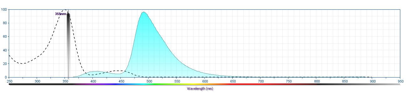
The antibody was conjugated to BD Horizon™ BUV496 which is part of the BD Horizon Brilliant™ Ultraviolet family of dyes. This dye is a tandem fluorochrome of BD Horizon BUV395 with an Ex Max of 348-nm and an acceptor dye with an Em Max at 496-nm. BD Horizon BUV496 can be excited by the ultraviolet laser (355 nm) and detected with a 515/30 nm filter with a 450LP. Due to the excitation of the acceptor dye by other laser lines, there may be significant spillover into the channel detecting BD Horizon V500 or BV510 (eg, 525/40-nm filter). However, the spillover can be corrected through compensation as with any other dye combination.
Development References (7)
-
Schubert J, Lanier LL, Schmidt RE. Cluster report: CD56. In: Knapp W. W. Knapp .. et al., ed. Leucocyte typing IV : white cell differentiation antigens. Oxford New York: Oxford University Press; 1989:699-702.
-
Edelman GM. Cell adhesion molecules.. Science. 1983; 219(4584):450-7. (Biology). View Reference
-
Hercend T, Griffin JD, Bensussan A, et al. Generation of monoclonal antibodies to a human natural killer clone. Characterization of two natural killer-associated antigens, NKH1A and NKH2, expressed on subsets of large granular lymphocytes.. J Clin Invest. 1985; 75(3):932-43. (Biology). View Reference
-
Lanier LL, Chang C, Azuma M, Ruitenberg JJ, Hemperly JJ, Phillips JH. Molecular and functional analysis of human natural killer cell-associated neural cell adhesion molecule (N-CAM/CD56). J Immunol. 1991; 146(12):4421-4426. (Clone-specific: Flow cytometry, Fluorescence activated cell sorting, Immunoprecipitation). View Reference
-
Lanier LL, Le AM, Civin CI, Loken MR, Phillips JH. The relationship of CD16 (Leu-11) and Leu-19 (NKH-1) antigen expression on human peripheral blood NK cells and cytotoxic T lymphocytes. J Immunol. 1986; 136(12):4480-4486. (Immunogen: Flow cytometry). View Reference
-
Lanier LL, Testi R, Bindl J, Phillips JH. Identity of Leu-19 (CD56) leukocyte differentiation antigen and neural cell adhesion molecule. J Exp Med. 1989; 169(6):2233-2238. (Clone-specific: Immunoprecipitation). View Reference
-
Phillips JH, Lanier LL. Dissection of the lymphokine-activated killer phenomenon: relative contribution of peripheral blood natural killer cells and T lymphocytes to cytolysis. J Exp Med. 1986; 164(3):814-825. (Clone-specific: Flow cytometry). View Reference
Please refer to Support Documents for Quality Certificates
Global - Refer to manufacturer's instructions for use and related User Manuals and Technical data sheets before using this products as described
Comparisons, where applicable, are made against older BD Technology, manual methods or are general performance claims. Comparisons are not made against non-BD technologies, unless otherwise noted.
For Research Use Only. Not for use in diagnostic or therapeutic procedures.