Skip to main content Skip to navigation
PE Mouse Anti-Human ILT1 (CD85h)
Two-parameter flow cytometric analysis of ILT1 (CD85h) expression on human peripheral blood leucocytes. Human whole blood was stained with either PE Mouse IgG2b, κ Isotype Control (Cat. No. 555058; Left Plot) or PE Mouse Anti-Human ILT1 (CD85h) antibody (Cat. No. 566577; Right Plot) at 1 µg/test. Erythrocytes were lysed with BD Pharm Lyse™ Lysing Buffer (Cat. No. 555899). Two-parameter flow cytometric contour plots showing the correlated expression of ILT1 (CD85h) [or Ig Isotype control staining] versus side light-scatter (SSC-A) signals were derived from gated events with the forward and side light-scattering characteristics of viable leucocyte populations. Flow cytometric analysis was performed using a BD LSRFortessa™ X-20 Cell Analyzer System. Data shown on this Technical Data Sheet are not lot specific.
Product Details
Down Arrow Up Arrow


BD Pharmingen™
LILRA2; ILT1; ILT-1; LILRA2; LIR7; LIR-7
Human (QC Testing)
Mouse BALB/c IgG2b, κ
Human ILT1 Transfected Cell Line
Flow cytometry (Routinely Tested)
0.2 mg/ml
AB_2739744
Aqueous buffered solution containing ≤0.09% sodium azide.
RUO


Preparation And Storage

Store undiluted at 4°C and protected from prolonged exposure to light. Do not freeze. The monoclonal antibody was purified from tissue culture supernatant or ascites by affinity chromatography. The antibody was conjugated with R-PE under optimum conditions, and unconjugated antibody and free PE were removed.

Product Notices

  1. Since applications vary, each investigator should titrate the reagent to obtain optimal results.
  2. An isotype control should be used at the same concentration as the antibody of interest.
  3. Caution: Sodium azide yields highly toxic hydrazoic acid under acidic conditions. Dilute azide compounds in running water before discarding to avoid accumulation of potentially explosive deposits in plumbing.
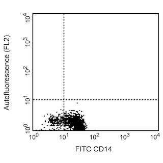
  4. For fluorochrome spectra and suitable instrument settings, please refer to our Multicolor Flow Cytometry web page at www.bdbiosciences.com/colors.
  5. Please refer to www.bdbiosciences.com/us/s/resources for technical protocols.
566577 Rev. 1
Antibody Details
Down Arrow Up Arrow
24/ILT1

The monoclonal antibody 24 specifically recognizes Immunoglobulin-like transcript 1 (ILT1) which is also known as Leukocyte immunoglobulin-like receptor 7 (LIR-7).  ILT1 (CD85h) is encoded by LILRA2 (Leukocyte immunoglobulin like receptor A2) and belongs to the Leukocyte immunoglobulin like receptors (LILR) gene family. This immunoreceptor is a ~69 kDa type I transmembrane glycoprotein that contains 4 Ig-like C2-type immunoglobulin-like extracellular domains and a short cytoplasmic domain. It is expressed on monocytes, macrophages, myeloid dendritic cells, and at lower levels on some granulocytes and a small subset of natural killer (NK) cells. ILT1 (CD85h) serves as an activating immunoreceptor through its short cytoplasmic domain that associates with the signal-transducing Fc receptor γ chain. This receptor complex mediates suppression of innate immune responses and inhibition of dendritic cell differentiation and antigen presentation. ILT1 (CD85h) may act as a receptor for immunoglobulins cleaved by microbial proteases.


566577 Rev. 1
Format Details
Down Arrow Up Arrow
PE
R-Phycoerythrin (PE), is part of the BD family of Phycobiliprotein dyes. This fluorochrome is a multimeric fluorescent phycobiliprotein with excitation maximum (Ex Max) of 496 nm and 566 nm and an emission maximum (Em Max) at 576 nm. PE is designed to be excited by the Blue (488 nm), Green (532 nm) and Yellow-Green (561 nm) lasers and detected using an optical filter centered near 575 nm (e.g., a 575/26-nm bandpass filter). As PE is excited by multiple lasers, this can result in cross-laser excitation and fluorescence spillover on instruments with various combinations of Blue, Green, and Yellow-Green lasers. Please ensure that your instrument’s configurations (lasers and optical filters) are appropriate for this dye.
altImg
PE
Yellow-Green 488 nm, 532 nm, 561 nm
496 nm, 566 nm
576 nm
566577 Rev.1
Citations & References
Down Arrow Up Arrow

Development References (5)

  1. Hirayasu K, Saito F, Suenaga T, et al. Microbially cleaved immunoglobulins are sensed by the innate immune receptor LILRA2. Nat Microbiol. 2016; 1(6):16054. (Biology). View Reference
  2. Lee DJ, Sieling PA, Ochoa MT, et al. LILRA2 activation inhibits dendritic cell differentiation and antigen presentation to T cells. J Immunol. 2007; 179(12):8128-8136. (Immunogen: Functional assay, Immunofluorescence). View Reference
  3. Lichterfeld M, Yu XG. The emerging role of leukocyte immunoglobulin-like receptors (LILRs) in HIV-1 infection. J Leukoc Biol. 2012; 91(1):27-33. (Biology). View Reference
  4. Nakajima H, Samaridis J, Angman L, Colonna M. Human myeloid cells express an activating ILT receptor (ILT1) that associates with Fc receptor gamma-chain.. J Immunol. 1999; 162(1):5-8. (Biology). View Reference
  5. Tedla N, Bandeira-Melo C, Tassinari P, et al. Activation of human eosinophils through leukocyte immunoglobulin-like receptor 7. Proc Natl Acad Sci U S A. 2003; 100(3):1174-1179. (Biology). View Reference
View All (5) View Less
566577 Rev. 1

Please refer to Support Documents for Quality Certificates


Global - Refer to manufacturer's instructions for use and related User Manuals and Technical data sheets before using this products as described


Comparisons, where applicable, are made against older BD Technology, manual methods or are general performance claims.  Comparisons are not made against non-BD technologies, unless otherwise noted.

For Research Use Only. Not for use in diagnostic or therapeutic procedures.