Skip to main content Skip to navigation
PE Mouse Anti-Human HLA-B7
Multiparameter flow cytometric analysis of HLA-B7 expression on human peripheral blood leucocyte populations from a HLA-B7-positive donor. Human whole blood from a HLA-B7-positive donor was stained with either PE Mouse IgG1, κ Isotype Control (Cat. No. 554680; Left Plot) or PE Mouse Anti-Human HLA-B7 antibody (Cat. No. 566654; Right Plot) at 1 µg/ml. Erythrocytes were lysed with BD Pharm Lyse™ Lysing Buffer (Cat. No. 555899). Two-parameter pseudocolor dot plots showing the correlated expression of HLA-B7 (or Ig Isotype control staining) versus side light-scatter (SSC-A) signals were derived from gated events with the forward and side light-scatter characteristics of viable leucocyte populations. Flow cytometric analysis was performed using a BD FACSCanto™ II Flow Cytometer System. Data shown on this Technical Data Sheet are not lot specific.
PE Mouse Anti-Human HLA-B7
Flow cytometric analysis of HLA-B7 expression on Jurkat cells. Jurkat cells (Acute T cell leukemia, ATCC TIB-152) were stained with either PE Mouse IgG1, κ Isotype Control (dashed line histogram) or PE Mouse Anti-Human HLA-B7 antibody (solid line histogram) at 1 µg/ml. Florescence histograms showing HLA-B7 expression (or Ig Isotype control staining) were derived from gated events with the forward and side light-scatter characteristics of viable cells. Flow cytometric analysis was performed using a BD FACSCanto™ II Flow Cytometer System. Data shown on this Technical Data Sheet are not lot specific.
Multiparameter flow cytometric analysis of HLA-B7 expression on human peripheral blood leucocyte populations from a HLA-B7-positive donor. Human whole blood from a HLA-B7-positive donor was stained with either PE Mouse IgG1, κ Isotype Control (Cat. No. 554680; Left Plot) or PE Mouse Anti-Human HLA-B7 antibody (Cat. No. 566654; Right Plot) at 1 µg/ml. Erythrocytes were lysed with BD Pharm Lyse™ Lysing Buffer (Cat. No. 555899). Two-parameter pseudocolor dot plots showing the correlated expression of HLA-B7 (or Ig Isotype control staining) versus side light-scatter (SSC-A) signals were derived from gated events with the forward and side light-scatter characteristics of viable leucocyte populations. Flow cytometric analysis was performed using a BD FACSCanto™ II Flow Cytometer System. Data shown on this Technical Data Sheet are not lot specific.
Flow cytometric analysis of HLA-B7 expression on Jurkat cells. Jurkat cells (Acute T cell leukemia, ATCC TIB-152) were stained with either PE Mouse IgG1, κ Isotype Control (dashed line histogram) or PE Mouse Anti-Human HLA-B7 antibody (solid line histogram) at 1 µg/ml. Florescence histograms showing HLA-B7 expression (or Ig Isotype control staining) were derived from gated events with the forward and side light-scatter characteristics of viable cells. Flow cytometric analysis was performed using a BD FACSCanto™ II Flow Cytometer System. Data shown on this Technical Data Sheet are not lot specific.
Product Details
Down Arrow Up Arrow


BD Pharmingen™
HLA-B*07; HLAB7; MHC class I B7; MHC class I antigen B7
Human (QC Testing)
Mouse IgG1, κ
Papain-solubilized Human HLA-B7
Flow cytometry (Routinely Tested)
0.2 mg/ml
AB_2869811
Aqueous buffered solution containing ≤0.09% sodium azide.
RUO


Preparation And Storage

Store undiluted at 4°C and protected from prolonged exposure to light. Do not freeze. The monoclonal antibody was purified from tissue culture supernatant or ascites by affinity chromatography. The antibody was conjugated with R-PE under optimum conditions, and unconjugated antibody and free PE were removed.

Product Notices

  1. Since applications vary, each investigator should titrate the reagent to obtain optimal results.
  2. An isotype control should be used at the same concentration as the antibody of interest.
  3. Caution: Sodium azide yields highly toxic hydrazoic acid under acidic conditions. Dilute azide compounds in running water before discarding to avoid accumulation of potentially explosive deposits in plumbing.
  4. For fluorochrome spectra and suitable instrument settings, please refer to our Multicolor Flow Cytometry web page at www.bdbiosciences.com/colors.
  5. Please refer to www.bdbiosciences.com/us/s/resources for technical protocols.
566654 Rev. 2
Antibody Details
Down Arrow Up Arrow
BB7.1

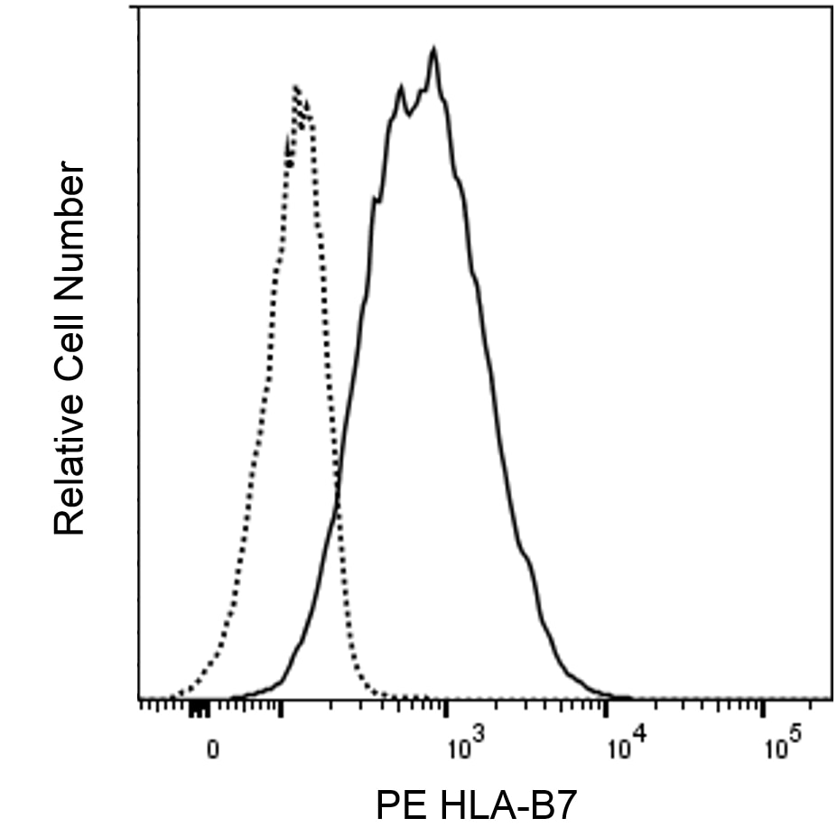
The BB7.1 monoclonal antibody specifically recognizes the Human Leukocyte Antigen-B7 (HLA-B7) alpha subunit. The HLA-B7 alpha chain can noncovalently heterodimerize with β2-microglobulin to form a Major Histocompatibility Complex (MHC) class I B7 antigen that is involved in antigen presentation to CD8+ T cells or might serve as an NK cell receptor ligand. HLA-B7 is a serotype that identifies the more common HLA-B (HLA-B*07) encoded gene products which are expressed on nearly all nucleated cells of HLA-B7-positive individuals. B7 is a risk factor for cervical cancer, sarcoidosis, and some spondylarthropathies. The BB7.1 antibody can be used to distinguish false HLA-B27-positve staining when used in conjunction with some HLA-B27 antibodies that crossreact with HLA-B7. Although highly specific, the BB7.1 antibody can reportedly crossreact with HLA-B42 and HLA-B81 antigens.


566654 Rev. 2
Format Details
Down Arrow Up Arrow
PE
R-Phycoerythrin (PE), is part of the BD family of Phycobiliprotein dyes. This fluorochrome is a multimeric fluorescent phycobiliprotein with excitation maximum (Ex Max) of 496 nm and 566 nm and an emission maximum (Em Max) at 576 nm. PE is designed to be excited by the Blue (488 nm), Green (532 nm) and Yellow-Green (561 nm) lasers and detected using an optical filter centered near 575 nm (e.g., a 575/26-nm bandpass filter). As PE is excited by multiple lasers, this can result in cross-laser excitation and fluorescence spillover on instruments with various combinations of Blue, Green, and Yellow-Green lasers. Please ensure that your instrument’s configurations (lasers and optical filters) are appropriate for this dye.
altImg
PE
Yellow-Green 488 nm, 532 nm, 561 nm
496 nm, 566 nm
576 nm
566654 Rev.2
Citations & References
Down Arrow Up Arrow

Development References (4)

  1. Brodsky FM, Parham P, Barnstable CJ, Crumpton MJ, Bodmer WF. Monoclonal antibodies for analysis of the HLA system. Immunol Rev. 1979; 47:3-6. (Immunogen: Immunoaffinity chromatography, Radioimmunoassay). View Reference
  2. Hilton HG, Parham P. Direct binding to antigen-coated beads refines the specificity and cross-reactivity of four monoclonal antibodies that recognize polymorphic epitopes of HLA class I molecules.. Tissue Antigens. 2013; 81(4):212-20. (Clone-specific: Cytometric Bead Array). View Reference
  3. Parham P. Purification of immunologically active HLA-A and -B antigens by a series of monoclonal antibody columns.. J Biol Chem. 1979; 254(18):8709-12. (Clone-specific: Immunoaffinity chromatography, Radioimmunoassay). View Reference
  4. Reches A, Nachmani D, Berhani O, et al. HNRNPR Regulates the Expression of Classical and Nonclassical MHC Class I Proteins.. J Immunol. 2016; 196(12):4967-76. (Clone-specific: Flow cytometry). View Reference
View All (4) View Less
566654 Rev. 2

Please refer to Support Documents for Quality Certificates


Global - Refer to manufacturer's instructions for use and related User Manuals and Technical data sheets before using this products as described


Comparisons, where applicable, are made against older BD Technology, manual methods or are general performance claims.  Comparisons are not made against non-BD technologies, unless otherwise noted.

For Research Use Only. Not for use in diagnostic or therapeutic procedures.