![APC Mouse Anti-Mouse I-A[b]](/content/dam/bdb/products/global/reagents/flow-cytometry-reagents/research-reagents/single-color-antibodies-ruo/562xxx/5628xx/562823_base/562823Image1.png)
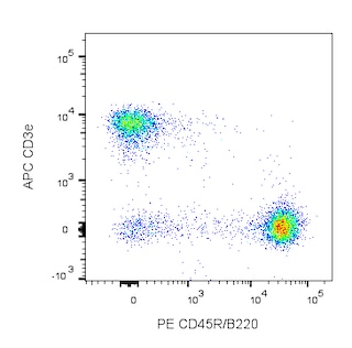
Multicolor flow cytometric analysis of I-A[b] expression on C57BL/6 mouse splenocytes. Splenic leucocytes were stained simultaneously with PE Rat anti-Mouse CD45R (Cat. No. 553090/553089/561878) and PE Rat anti-Mouse CD11b (Cat. No. 553311/557397/561689) antibodies and with either APC Mouse IgG2a, κ Isotype Control (Cat. No. 550882; Left Panel) or APC Mouse anti-Mouse I-A[b] antibody (Cat. No. 562823; Right Panel). Two-color flow cytometric dot plots show the correlated expression patterns of I-A[b] (or Ig Isotype control staining) versus CD45R and CD11b for gated events with the forward and side light-scatter characteristics of viable splenic leucocytes. Flow cytometry was performed using a BD™ LSR II Flow Cytometer System.
Multicolor flow cytometric analysis of I-A[b] expression on C57BL/6 mouse splenocytes. Splenic leucocytes were stained simultaneously with PE Rat anti-Mouse CD45R (Cat. No. 553090/553089/561878) and PE Rat anti-Mouse CD11b (Cat. No. 553311/557397/561689) antibodies and with either APC Mouse IgG2a, κ Isotype Control (Cat. No. 550882; Left Panel) or APC Mouse anti-Mouse I-A[b] antibody (Cat. No. 562823; Right Panel). Two-color flow cytometric dot plots show the correlated expression patterns of I-A[b] (or Ig Isotype control staining) versus CD45R and CD11b for gated events with the forward and side light-scatter characteristics of viable splenic leucocytes. Flow cytometry was performed using a BD™ LSR II Flow Cytometer System.
Product Details


BD Pharmingen™
Mouse MHC/H2 class II histocompatibility antigen, I-Ab
Mouse (QC Testing)
Mouse BALB/c IgG2a, κ
C57BL/10J Mouse Splenocytes
Flow cytometry (Routinely Tested)
0.2 mg/ml
AB_2737818
Aqueous buffered solution containing ≤0.09% sodium azide.
RUO
Preparation And Storage
Store undiluted at 4°C and protected from prolonged exposure to light. Do not freeze. The monoclonal antibody was purified from tissue culture supernatant or ascites by affinity chromatography. The antibody was conjugated to APC under optimum conditions, and unconjugated antibody and free APC were removed.
Product Notices
- Since applications vary, each investigator should titrate the reagent to obtain optimal results.
- An isotype control should be used at the same concentration as the antibody of interest.
- Please refer to www.bdbiosciences.com/us/s/resources for technical protocols.
- Caution: Sodium azide yields highly toxic hydrazoic acid under acidic conditions. Dilute azide compounds in running water before discarding to avoid accumulation of potentially explosive deposits in plumbing.
- This APC-conjugated reagent can be used in any flow cytometer equipped with a dye, HeNe, or red diode laser.
- For fluorochrome spectra and suitable instrument settings, please refer to our Multicolor Flow Cytometry web page at www.bdbiosciences.com/colors.
Companion Products
Stain Buffer (FBS) RUO
Size
500 mL
Cat No.
554656

Lysing Buffer RUO
Size
100 mL
Cat No.
555899

APC Mouse IgG2a κ Isotype Control RUO
Size
0.1 mg
Cat No.
550882
.png?imwidth=320)
PE Rat Anti-Mouse CD45R/B220 RUO
Size
0.2 mg
Cat No.
553090
PE Rat Anti-Mouse CD45R/B220 RUO
Size
0.1 mg
Cat No.
553089
PE Rat Anti-Mouse CD45R/B220 RUO
Size
25 µg
Cat No.
561878
562823 Rev. 1
Antibody Details
AF6-120.1
The AF6-120.1 antibody reognizes the I-A[b] MHC class II alloantigen. It crossreacts with cells from mice of the H-2[k] and H-2[u] haplotypes. Reactivity with other haplotypes (e.g., d, f, g7, p, q, r, s) has not been observed.
562823 Rev. 1
Format Details
APC
Allophycocyanin (APC), is part of the BD family of phycobiliprotein dyes. This fluorochrome is a multimeric fluorescent phycobiliprotein with excitation maximum (Ex Max) of 651 nm and an emission maximum (Em Max) at 660 nm. APC is designed to be excited by the Red (627-640 nm) laser and detected using an optical filter centered near 660 nm (e.g., a 660/20 nm bandpass filter). Please ensure that your instrument’s configurations (lasers and optical filters) are appropriate for this dye.

APC
Red 627-640 nm
651 nm
660 nm
562823 Rev.1
Citations & References
Development References (7)
-
BD Biosciences Pharmingen. Unpublished results. .
-
Beck BN, Buerstedde JM, Krco CJ, Nilson AE, Chase CG, McKean DJ. Characterization of cell lines expressing mutant I-Ab and I-Ak molecules allows the definition of distinct serologic epitopes on A alpha and A beta polypeptides. J Immunol. 1986; 136(8):2953-2961. (Clone-specific: Flow cytometry). View Reference
-
Cohn LE, Glimcher LH, Waldmann RA, et al. Identification of functional regions on the I-Ab molecule by site-directed mutagenesis. Proc Natl Acad Sci U S A. 1986; 83(3):747-751. (Clone-specific: Flow cytometry). View Reference
-
Hattori M, Buse JB, Jackson RA, et al. The NOD mouse: recessive diabetogenic gene in the major histocompatibility complex. Science. 1986; 231(4739):733-735. (Biology). View Reference
-
Nabozny GH, Baisch JM, Cheng S, et al. HLA-DQ8 transgenic mice are highly susceptible to collagen-induced arthritis: a novel model for human polyarthritis. J Exp Med. 1996; 183(1):27-37. (Clone-specific: Flow cytometry). View Reference
-
Stall AM. Personal Communication. .
-
Wall KA, Lorber MI, Loken MR, McClatchey S, Fitch FW. Inhibition of proliferation of MIs- and Ia-reactive cloned T cells by a monoclonal antibody against a determinant shared by I-A and I-E.. J Immunol. 1983; 131(3):1056-64. (Clone-specific: Flow cytometry). View Reference
562823 Rev. 1
Please refer to Support Documents for Quality Certificates
Global - Refer to manufacturer's instructions for use and related User Manuals and Technical data sheets before using this products as described
Comparisons, where applicable, are made against older BD Technology, manual methods or are general performance claims. Comparisons are not made against non-BD technologies, unless otherwise noted.
For Research Use Only. Not for use in diagnostic or therapeutic procedures.