Old Browser
This page has been recently translated and is available in French now.
Looks like you're visiting us from {countryName}.
Would you like to stay on the current country site or be switched to your country?


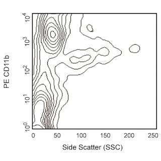
Three-color flow cytometric analysis of F4/80-Like Receptor expression on mouse bone marrow cells. Mouse bone-marrow cells were preincubated with Purified Rat Anti-Mouse CD16/CD32 antibody (Mouse BD Fc Block™) (Cat. No. 553141/553142). The cells were then stained with PE Rat Anti-Mouse CD11b (Cat. No. 5553311/557397/561689) and FITC Hamster Anti-Mouse CD11c (Cat. No. 553801/557400/561045) antibodies and either Alexa Fluor® 647 Rat IgG2a, κ Isotype Control (Cat. No. 557690, Left Panels) or Alexa Fluor® 647 Rat Anti-Mouse F4/80-Like Receptor antibody (Cat. No. 564226; Right Panels). Two-color flow cytometric dot plots showing the correlated expression patterns of CD11b (Upper Panels) or CD11c (Lower Panels) versus F4/80-Like Receptor (or Ig Isotype control staining) were derived from gated events with the forward and side light-scatter characteristics of viable bone marrow cells. Flow cytometric analysis was performed using a BD LSRFortessa™ Cell Analyzer System.


BD Pharmingen™ Alexa Fluor® 647 Rat Anti-Mouse F4/80-Like Receptor

Regulatory Status Legend
Any use of products other than the permitted use without the express written authorization of Becton, Dickinson and Company is strictly prohibited.
Preparation And Storage
Product Notices
- Since applications vary, each investigator should titrate the reagent to obtain optimal results.
- An isotype control should be used at the same concentration as the antibody of interest.
- Caution: Sodium azide yields highly toxic hydrazoic acid under acidic conditions. Dilute azide compounds in running water before discarding to avoid accumulation of potentially explosive deposits in plumbing.
- The Alexa Fluor®, Pacific Blue™, and Cascade Blue® dye antibody conjugates in this product are sold under license from Molecular Probes, Inc. for research use only, excluding use in combination with microarrays, or as analyte specific reagents. The Alexa Fluor® dyes (except for Alexa Fluor® 430), Pacific Blue™ dye, and Cascade Blue® dye are covered by pending and issued patents.
- Alexa Fluor® is a registered trademark of Molecular Probes, Inc., Eugene, OR.
- Alexa Fluor® 647 fluorochrome emission is collected at the same instrument settings as for allophycocyanin (APC).
- For fluorochrome spectra and suitable instrument settings, please refer to our Multicolor Flow Cytometry web page at www.bdbiosciences.com/colors.
- Please refer to www.bdbiosciences.com/us/s/resources for technical protocols.
Companion Products


The 6F12 antibody reacts with a 7-transmembrane-domain protein, which is similar to the F4/80 macrophage antigen of the EGF-TM7 protein family and is encoded by the Emr4 gene. The FIRE protein is expressed on myeloid cells with a denditic cell (DC) developmental potential, including subsets of DC and macrophages in the spleen and lymph nodes, most resident peritoneal macrophages, many peripheral blood monocytes, and a subpopulation of bone-marrow myeloid-cell progenitors. The protein is not detected on peripheral T and B lymphocytes, and it is down-regulated on thioglycollate-elicited peritoneal macrophages and on dendritic cells activated by GM-CSF, IFN-γ, anti-CD40, and LPS. Using soluble biotinylated fusion protein, a FIRE ligand was detected on a mouse IgG+ B lymphoma cell line (A20), but not on myeloid, fibroblast, or T-cell lines, suggesting that the FIRE protein may be involved in immunoregulatory interactions between antigen-presenting cells and B lymphocytes.
Development References (2)
-
Caminschi I, Lucas KM, O'Keeffe MA, et al. Molecular cloning of F4/80-like-receptor, a seven-span membrane protein expressed differentially by dendritic cell and monocyte-macrophage subpopulations. J Immunol. 2001; 167(7):3570-3576. (Immunogen: Immunohistochemistry, Immunoprecipitation, Western blot). View Reference
-
Stacey M, Chang GW, Sanos SL, et al. EMR4, a novel epidermal growth factor (EGF)-TM7 molecule up-regulated in activated mouse macrophages, binds to a putative cellular ligand on B lymphoma cell line A20. J Biol Chem. 2002; 277(32):29283. (Biology). View Reference
Please refer to Support Documents for Quality Certificates
Global - Refer to manufacturer's instructions for use and related User Manuals and Technical data sheets before using this products as described
Comparisons, where applicable, are made against older BD Technology, manual methods or are general performance claims. Comparisons are not made against non-BD technologies, unless otherwise noted.
For Research Use Only. Not for use in diagnostic or therapeutic procedures.
Report a Site Issue
This form is intended to help us improve our website experience. For other support, please visit our Contact Us page.