Skip to main content Skip to navigation
Alexa Fluor® 647 Mouse Anti-Human CD45RC
Multiparameter flow cytometric analysis of CD45RC expression on human peripheral blood leucocytes.       Human whole blood was stained with PerCP-Cy™5.5 Mouse Anti-Human CD4 (Cat. No. 560650) and BD Horizon™ BV421 Mouse Anti-Human CD19 (Cat. No. 562440/562441) antibodies, and either Alexa Fluor® 647 Mouse IgG1 κ Isotype Control (Cat. No. 557714; Left Plots) or Alexa Fluor® 647 Mouse Anti-Human CD45RC antibody (Cat. No. 565857; Right Plots). Erythrocytes were lysed with BD Pharm Lyse™ Lysing Buffer (Cat. No. 555899).      Left Panel - Two-parameter flow cytometric contour plots showing the correlated expression of CD45RC (or Ig Isotype control staining) versus side light-scatter (SSC-A) signals were derived from gated events with the forward and side light-scatter characteristics of viable leucocyte populations.      Middle and Right Panels - Two-color flow cytometric contour plots showing the correlated expression of CD4 (Middle Panel) or CD19 (Right Panel) versus CD45RC (or Ig Isotype control staining) were derived from gated events with the forward and side light-scatter characteristics of viable lymphocytes.      Flow cytometric analysis was performed using a BD FACSCanto™ II Flow Cytometer System.
Product Details
Down Arrow Up Arrow


BD Pharmingen™
PTPRC; CD45R; L-CA; LCA; Leukocyte common antigen; LY5; T200
Human (QC Testing)
Mouse BALB/c IgG1, κ
Lymph node cells from human donor with B-CLL
Flow cytometry (Routinely Tested)
5 µl
IV N913
AB_2869721
Aqueous buffered solution containing BSA and ≤0.09% sodium azide.
RUO


Preparation And Storage

Store undiluted at 4°C and protected from prolonged exposure to light. Do not freeze. The monoclonal antibody was purified from tissue culture supernatant or ascites by affinity chromatography. The antibody was conjugated to Alexa Fluor® 647 under optimum conditions, and unreacted Alexa Fluor® 647 was removed.

Product Notices

  1. This reagent has been pre-diluted for use at the recommended Volume per Test. We typically use 1 × 10^6 cells in a 100-µl experimental sample (a test).
  2. Please refer to www.bdbiosciences.com/us/s/resources for technical protocols.
  3. The Alexa Fluor®, Pacific Blue™, and Cascade Blue® dye antibody conjugates in this product are sold under license from Molecular Probes, Inc. for research use only, excluding use in combination with microarrays, or as analyte specific reagents. The Alexa Fluor® dyes (except for Alexa Fluor® 430), Pacific Blue™ dye, and Cascade Blue® dye are covered by pending and issued patents.
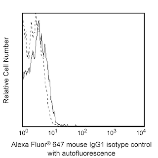
  4. Alexa Fluor® 647 fluorochrome emission is collected at the same instrument settings as for allophycocyanin (APC).
  5. Alexa Fluor® is a registered trademark of Molecular Probes, Inc., Eugene, OR.
  6. Cy is a trademark of Amersham Biosciences Limited.
  7. Source of all serum proteins is from USDA inspected abattoirs located in the United States.
  8. Caution: Sodium azide yields highly toxic hydrazoic acid under acidic conditions. Dilute azide compounds in running water before discarding to avoid accumulation of potentially explosive deposits in plumbing.
  9. For fluorochrome spectra and suitable instrument settings, please refer to our Multicolor Flow Cytometry web page at www.bdbiosciences.com/colors.
  10. An isotype control should be used at the same concentration as the antibody of interest.
565857 Rev. 1
Antibody Details
Down Arrow Up Arrow
MT2

The MT2 monoclonal antibody specifically binds to CD45RC isoforms of CD45 that share exon 6-encoded sequences. CD45 is a type I transmembrane glycoprotein that serves as a receptor-like protein tyrosine phosphatase (PTP). CD45RC is highly expressed on medullary thymocytes, CD8+ T cells, B cells, NK cells, and by subsets of cortical thymocytes or CD4+ T cells. CD45RC may play a role in regulating receptor-mediated leucocyte signaling responses.

565857 Rev. 1
Format Details
Down Arrow Up Arrow
Alexa Fluor™ 647
Alexa Fluor™ 647 Dye is part of the BD red family of dyes. This is a small organic fluorochrome with an excitation maximum (Ex Max) at 653-nm and an emission maximum (Em Max) at 669-nm. Alexa Fluor 647 is designed to be excited by the Red laser (627-640 nm) and detected using an optical filter centered near 520-nm (e.g., a 660/20 nm bandpass filter). Please ensure that your instrument’s configurations (lasers and optical filters) are appropriate for this dye.
Alexa Fluor™ 647
Red 627-640 nm
653 nm
669 nm
565857 Rev.1
Citations & References
Down Arrow Up Arrow

Development References (6)

  1. Grotjahn C, Stein H, Hadam MR. Monoclonal antibodies OTH75E4 (6w N121) and MT2 are both assigned to CD45RC. Tissue Antigens. 1996; 48(4):446. (Clone-specific: Blocking, Immunohistochemistry). View Reference
  2. Ordonez L, Bernard I, L'faqihi-Olive FE, Tervaert JW, Damoiseaux J, Saoudi A. CD45RC isoform expression identifies functionally distinct T cell subsets differentially distributed between healthy individuals and AAV patients.. PLoS ONE. 2009; 4(4):e5287. (Clone-specific: Cell separation, Flow cytometry, Fluorescence activated cell sorting). View Reference
  3. Poppema S, Hollema H, Visser L, Vos H. Monoclonal antibodies (MT1, MT2, MB1, MB2, MB3) reactive with leukocyte subsets in paraffin-embedded tissue sections.. Am J Pathol. 1987; 127(3):418-29. (Immunogen: Immunohistochemistry, Western blot). View Reference
  4. Poppema S, Lai R, Visser L, Yan XJ. CD45 (leucocyte common antigen) expression in T and B lymphocyte subsets.. Leuk Lymphoma. 1996; 20(3-4):217-22. (Clone-specific). View Reference
  5. Schwinzer R. Cluster Report: CD45/CD45R. In: Knapp W. W. Knapp .. et al., ed. Leucocyte typing IV : white cell differentiation antigens. Oxford New York: Oxford University Press; 1989:628-634.
  6. Zapata JM, Pulido R, Acevedo A, Sánchez-Madrid F, de Landázuri MO. Human CD45RC specificity. A novel marker for T cells at different maturation and activation stages.. J Immunol. 1994; 152(8):3852-61. (Biology). View Reference
View All (6) View Less
565857 Rev. 1

Please refer to Support Documents for Quality Certificates


Global - Refer to manufacturer's instructions for use and related User Manuals and Technical data sheets before using this products as described


Comparisons, where applicable, are made against older BD Technology, manual methods or are general performance claims.  Comparisons are not made against non-BD technologies, unless otherwise noted.

For Research Use Only. Not for use in diagnostic or therapeutic procedures.