Old Browser
This page has been recently translated and is available in French now.
Looks like you're visiting us from {countryName}.
Would you like to stay on the current country site or be switched to your country?


Immunohistochemistry analysis of CD88 expression on human tonsil paraffin sections. Dendritic cells (Left Panel) or granulocytes (Right Panel) from human tonsil sections were stained with Purified Rabbit Anti-Human CD88 (2.5 µg/ml; Cat. No. 550733), followed by Biotinylated Donkey Anti-Rabbit Ig (5 µg/ml; Jackson ImmunoResearch Cat. No. 711-065-152) and Streptavidin HRP (Cat. No. 550946).

Flow cytometric analysis of CD88 expression on human granulocytes. Whole blood was stained with either Purified Rabbit Anti-Human CD88 (solid line histogram) or Purified Rabbit IgG (Jackson ImmunoResearch Cat. No. 011-000-003; dashed line histogram) at 1 µg/test, followed by FITC Goat Anti-Rabbit IgG (Cat. No. 554020). Erythrocytes were lysed with BD Pharm Lyse™ Lysing Buffer (Cat. No. 349202). Fluorescent histograms depicting CD88 (or Ig isotype control) expression were derived from gated events with the side and forward light-scattering characteristics of viable granulocytes.


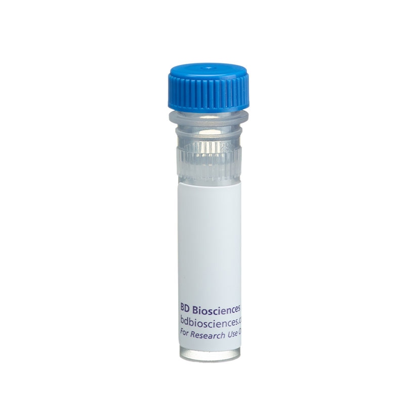
BD Pharmingen™ Purified Rabbit Anti-Human CD88

BD Pharmingen™ Purified Rabbit Anti-Human CD88

Regulatory Status Legend
Any use of products other than the permitted use without the express written authorization of Becton, Dickinson and Company is strictly prohibited.
Preparation And Storage
Recommended Assay Procedures
Immunohistochemistry: The purified C85-2506 clone can be used for immunohistochemistry to detect expression of the human C5aR. The antibody has been tested on frozen and fixation of paraffin sections of human tonsil and paraffin sections of human spleen, liver, colon and lung. The dilution range recommended for this antibody is 1-10 to 1-20. The C85-2506 clone does not cross react with C5aR expressed on mouse, rat, pig or monkey tissue. Using biotinylated donkey anti-rabbit Ig (Jackson ImmunoResearch) as the secondary antibody, followed by a peroxidase-based detection system, the expression of C5aR was observed on dendritic cells, granulocytes and monocytes of human tonsil.
Product Notices
- Since applications vary, each investigator should titrate the reagent to obtain optimal results.
- An isotype control should be used at the same concentration as the antibody of interest.
- Caution: Sodium azide yields highly toxic hydrazoic acid under acidic conditions. Dilute azide compounds in running water before discarding to avoid accumulation of potentially explosive deposits in plumbing.
- Source of all serum proteins is from USDA inspected abattoirs located in the United States.
- Sodium azide is a reversible inhibitor of oxidative metabolism; therefore, antibody preparations containing this preservative agent must not be used in cell cultures nor injected into animals. Sodium azide may be removed by washing stained cells or plate-bound antibody or dialyzing soluble antibody in sodium azide-free buffer. Since endotoxin may also affect the results of functional studies, we recommend the NA/LE (No Azide/Low Endotoxin) antibody format, if available, for in vitro and in vivo use.
- Please refer to http://regdocs.bd.com to access safety data sheets (SDS).
- Please refer to www.bdbiosciences.com/us/s/resources for technical protocols.
The human C5a receptor (C5aR) is expressed on granulocytes, monocytes, macrophages mast cells human cell lines of myeloid origin, and certain cell types in several organs, including dendritic cells of secondary lymphoid organs, liver hepatocytes, lung bronchial and alveolar epithelial cells, lung vascular smooth muscle, endothelial cells astrocytes, microglia, and fibroblast-like cells of the brain. The C85-2506 is a rabbit monoclonal antibody generated by fusion of splenocytes from rabbits immunized with human C5aR peptides and the rabbit fusion partner 240E-1.
Development References (10)
-
Fayyazi A, Sandau R, Duong LQ. C5a receptor and interleukin-6 are expressed in tissue macrophages and stimulated keratinocytes but not in pulmonary and intestinal epithelial cells. Am J Pathol. 1999; 154(2):495-501. (Biology). View Reference
-
Füreder W, Agis H, Willheim M. Differential expression of complement receptors on human basophils and mast cells. Evidence for mast cell heterogeneity and CD88/C5aR expression on skin mast cells. J Immunol. 1995; 155(6):3152-3160. (Biology). View Reference
-
Gasque P, Singhrao SK, Neal JW, Götze O, Morgan BP. Expression of the receptor for complement C5a (CD88) is up-regulated on reactive astrocytes, microglia, and endothelial cells in the inflamed human central nervous system. Am J Pathol. 1997; 150(1):31-41. (Biology). View Reference
-
Haviland DL, McCoy RL, Whitehead WT, et al. Cellular expression of the C5a anaphylatoxin receptor (C5aR): Demonstration of C5aR on nonmyeloid cells of the liver and lung. J Immunol. 1995; 154:1861-1869. (Biology).
-
Morelli A, Larregina A, Chuluyán I, Kolkowski E, Fainboim L. Expression and modulation of C5a receptor (CD88) on skin dendritic cells. Chemotactic effect of C5a on skin migratory dendritic cells. Immunology. 1996; 89(1):126-134. (Biology). View Reference
-
Nataf S, Davoust N, Barnum SR. Kinetics of anaphylatoxin C5a receptor expression during experimental allergic encephalomyelitis. J Neuroimmunol. 1998; 91(1-2):147-155. (Biology). View Reference
-
Paradisis PM, Campbell IL, Barnum SR. Elevated complement C5a receptor expression on neurons and glia in astrocyte-targeted interleukin-3 transgenic mice. Glia. 1998; 24(3):338-345. (Biology). View Reference
-
Spieker-Polet H, Sethupathi P, Yam PC, Knight KL. Rabbit monoclonal antibodies: generating a fusion partner to produce rabbit-rabbit hybridomas. Proc Natl Acad Sci U S A. 1995; 92(20):9348-9352. (Immunogen). View Reference
-
Stahel PF, Frei K, Eugster HP. TNF-alpha-mediated expression of the receptor for anaphylatoxin C5a on neurons in experimental Listeria meningoencephalitis. J Immunol. 1997; 159(2):861-869. (Biology). View Reference
-
Wetsel RA. Expression of the complement C5a anaphylatoxin receptor (C5aR) on non-myeloid cells. Immunol Lett. 1995; 44(2-3):183-187. (Biology). View Reference
Please refer to Support Documents for Quality Certificates
Global - Refer to manufacturer's instructions for use and related User Manuals and Technical data sheets before using this products as described
Comparisons, where applicable, are made against older BD Technology, manual methods or are general performance claims. Comparisons are not made against non-BD technologies, unless otherwise noted.
For Research Use Only. Not for use in diagnostic or therapeutic procedures.
Report a Site Issue
This form is intended to help us improve our website experience. For other support, please visit our Contact Us page.