BD Pharmingen™ APO-DIRECT™ Kit
(RUO)


Description
One of the later steps in apoptosis is DNA fragmentation, a process which results from the activation of endonucleases during the apoptotic program. These nucleases degrade the higher order chromatin structure into fragments of ~300 kb and subsequently into smaller DNA pieces of about 50 bp in length. A method which is often used to detect fragmented DNA utilizes a reaction catalyzed by exogenous TdT, often referred to as "end-labeling" or "TUNEL" (terminal deoxynucleotidyltransferase dUTP nick end labeling). The APO-DIRECT™ assay (Cat. No. 6536KK) is a single-step method for labeling DNA breaks with FITC-dUTP, followed by flow cytometric analysis. In the APO-BRDU™ assay, TdT catalyzes a template-independent addition of bromolated deoxyuridine triphosphates (Br-dUTP) to the 3'-hydroxyl (OH) termini of double- and single-stranded DNA. After incorporation, these sites are identified by flow cytometric means by staining the cells with a FITC-labeled anti-BrdU mAb.
APO-DIRECT™ is a single-step staining method for labeling DNA breaks to detect apoptotic cells by flow cytometry. Sufficient reagents are provided to process 50 tests. The kit includes 5 ml of positive and 5 ml of negative control cell suspensions, approximately 1 x 10^6 cells per ml in 70 % ( v/v) ethanol. The control cells are derived from a human lymphoma cell line and have been fixed as described below in the Fixation Protocol.
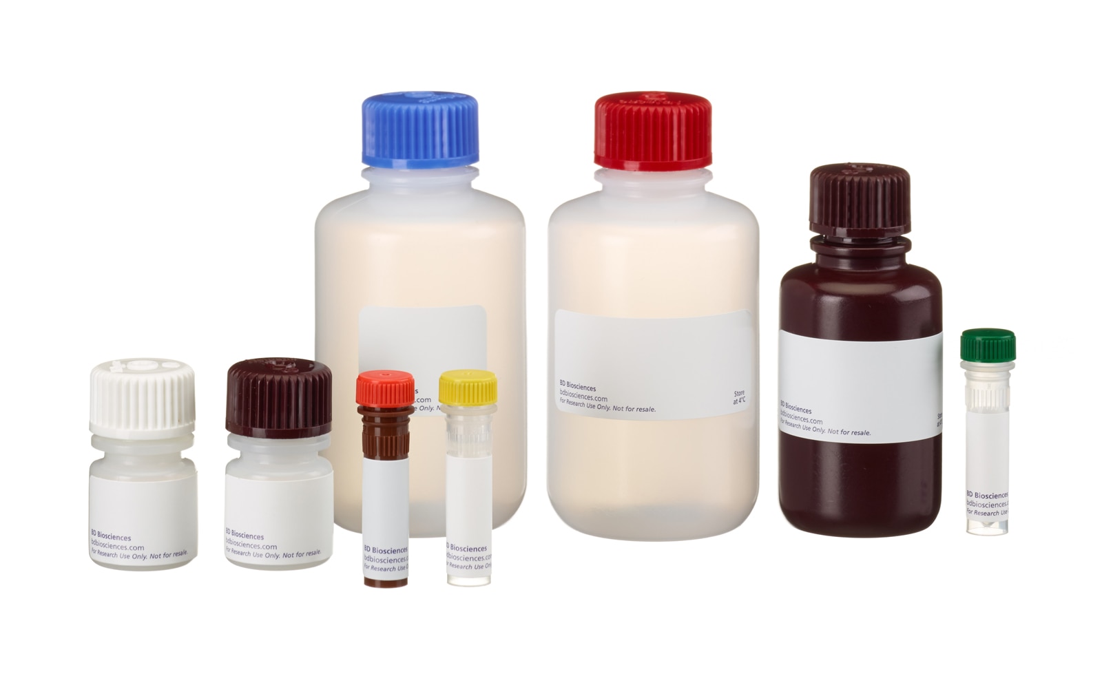
The APO-DIRECT™ Kit consists of two parts (part A and B).
Part A (Comp. No. 51-6536AK) consists of:
One 25 ml bottle of PI/RNase Staining Buffer (5 µg/ml, 200 µg/ml RNase) [Comp. No. 51-6551AZ],
One 0.5 ml vial of Reaction Buffer (containing cacodylic acid (dimethylarsenic)) [Comp. No. 51-6549AZ ] ,
One 100 ml bottle of Rinsing Buffer (containing 0.05% sodium azide) [Comp. No. 51-6550AZ]
One 100 ml botle of Wash Buffer (containing 0.05% sodium azide) [Comp. No. 51-6548AZ].
Part B (Comp. No. 51-6536BK) consists of:
One 0.4 ml bottle of FITC-dUTP (0.25 nMol) [Comp. No. 51-6555EZ]
One 5 ml bottle of Negative Control Cells [contains 70% (v/v) ethanol] [Comp. No. 51-6553LZ]
One 5 ml bottle of Positive Control Cells [contains 70% (v/v) ethanol] [Comp. No. 51-6552LZ]
One 0.038 ml bottle of TdT Enzyme [200 µg/ml (S.A.= 100,000 U/mg) in 50% (v/v) glycerol solution; will not freeze at -20°C] [Comp. No. 51-6554EZ].
Preparation And Storage
The APO-DIRECT™ Kit consists of two parts (A and B). Part A (Comp. No. 51-6536AK) is shipped at ambient temperature and should be stored at 4°C upon arrival. Part B (Comp. No. 51-6536BK) is shipped at –80°C on dry ice, and should be stored at –20°C upon arrival. BD Biosciences Pharmingen has determined that this shipping method is adequate to maintain the integrity of the kit components
Recommended Assay Procedures
METHODS FOR FIXATION, STAINING, AND ANALYZING APO-DIRECT™ SAMPLES
Fixation Protocol
This is a standard protocol used at BD Biosciences Pharmingen for quality control testing of the APO-DIRECT™ Kit. Cell fixation using paraformaldehyde is a required step in the APO-DIRECT™ assay. The following cell fixation procedure is a suggested method. Variables such as cell origin and growth conditions can affect the results. The fixation conditions provided below should be considered as guidelines. Additional experimentation may be required to obtain results comparable to the control cells provided with this kit. The positive and negative control cells provided in the APODIRECT™ Kit are already fixed.
1. Suspend the cells in 1% (w/v) paraformaldehyde in PBS (pH 7.4) at a concentration of 1-2 x 10^6 cells/ml.
2. Place the cell suspension on ice for 30-60 minutes.
3. Centrifuge cells for 5 min at 300 x g and discard the supernatant.
4. Wash the cells in 5 ml of PBS, then pellet the cells by centrifugation. Repeat the wash and centrifugation.
5. Resuspend the cell pellet in the residual PBS in the tube by gently vortexing the tube.
6. Adjust the cell concentration to 1-2 x 10^6 cells/ml in 70% (v/v) ice cold ethanol. Let cells stand for a minumum of 30 minutes on ice or in the freezer. See note below.
7. Store cells in 70% (v/v) ethanol at -20°C until use. Cells can be stored at -20°C several days before use.
Note: In some biological systems, storage of the cells at -20°C in 70% (v/v) ethanol for at least 12-18 hr prior to staining for apoptosis detection yields the best results. Cells can be stored at -20°C for several months before use.
Staining Protocol
The following protocol describes the method for measuring apoptosis in the positive and negative control cells that are provided in the APO-DIRECT™ Kit. The same procedure should be employed for measuring apoptosis in the cell specimens provided by the researcher.
1. Resuspend the positive (51-6552LZ) and negative (51-6553LZ) control cells by swirling the vials. Remove 1 ml aliquots of the control cell suspensions (approximately 1 x 10^6 cells/ml) and place in 12 x 75 mm centrifuge tubes. Centrifuge the control cell suspensions for 5 min at 300 x g and remove the 70% (v/v) ethanol by aspiration, being careful to not disturb the cell pellet.
2. Resuspend each tube of control cells with 1.0 ml of Wash Buffer (51-6548AZ) for each tube. Centrifuge as before and remove the supernatant by aspiration.
3. Repeat the Wash Buffer treatment (Step 2).
4. Resuspend each tube of the control cell pellets in 50 µl of the DNA Labeling Solution (prepared as described below).
For one assay, use the following amounts of DNA Labeling Solutions:
DNA Labeling Solution 1 Assay 6 Assays 12 Assays
Reaction Buffer (green cap) 10.00 μl 60.00 μl 120.00 μl
TdT Enzyme (yellow cap) 0.75 μl 4.50 μl 9.00 μl
FITC dUTP (orange cap) 8.00 μl 48.00 μl 96.00 μl
Distilled H2O 32.25 μl 193.50 μl 387.00 μl
Total Volume 51.00 μl 306.00 μl 612.00 μl
5. Incubate the cells in the DNA Labeling Solution for 60 min at 37°C. The reaction can also be carried out at RT overnight for the control cells. For test samples, the 60 min incubation time at 37°C may need to be adjusted to longer periods of time.
6. At the end of the incubation time, add 1.0 ml of Rinse Buffer (51-6550AZ) to each tube and centrifuge each tube at 300 x g for 5 min. Remove the supernatant by aspiration.
7. Repeat the cell rinsing with 1.0 ml of the Rinse Buffer, centrifuge, and remove the supernatant by aspiration.
8. Resuspend the cell pellet in 0.5 ml of the PI/RNase Staining Buffer (51-6551AZ).
Note: If the cell density is low, decrease the amount of PI/RNase Staining Buffer to 0.3 ml.
9. Incubate the cells in the dark for 30 min at RT.
10. Analyze the cells in PI/RNase solution by flow cytometry.
11. Analyze the cells within 3 hr of staining. Cells may begin to deteriorate if left overnight before analysis.
Analyzing APO-DIRECT™ Samples by Flow Cytometry
This assay is run on a flow cytometer equipped with a 488 nm Argon laser as the light source. Expected results using the positive and negative control cells are shown in Figs. 1 and 2. Two dyes are used; PI stains total DNA and FITC-dUTP stains apoptotic cells. PI fluoresces at about 623 nm and FITC at 520 nm. Two dual parameter and two single parameter displays are created with the flow cytometer data acquisition software. The gating display should be the standard dual parameter DNA doublet discrimination display with the DNA Area signal on the Y-axis and the DNA Width on the X-axis (Fig. 1). From this display, a gate is drawn around the non-clumped cells and the second gated dual parameter display is generated. The normal convention of this display is to put DNA (Linear Red Fluorescence) on the X-axis and the FITCdUTP (Log Green Fluorescence) on the Y-axis. Two single parameter-gated histograms, DNA and FITC-dUTP, can also be added but are not necessary. By using the dual parameter display method, not only are apoptotic cells resolved but their cell cycle stage is also determined.
TECHNICAL TIPS AND FREQUENTLY ASKED QUESTIONS
1. For researchers using adherent cell line systems, the cells in the supernatant have a higher probability of being apoptotic than do the adherent cells. Save cells in the supernatant for use in the assay prior to trypsinization of the adherent cell layer.
2. Cell fixation using a DNA cross-linking chemical fixative (e.g. paraformaldehyde) is an important step in analyzing apoptosis. Unfixed cells may lose smaller fragments of DNA that are not chemically fixed in place inside the cell prior to washing steps. The researcher may have to explore alternative fixation and permeabilization methods to fully exploit their systems.
3. A cytospin or centrifugal cytology slide can be prepared from the APO-DIRECT™ sample in the following manner: After completion of the FITC-dUTP staining, but prior to the PI/RNase treatment, put a drop of the stained cells on a slide, spin it and observe the sample under a fluorescence microscope.
4. To minimize cell loss during the assay, we recommend the use of 12 x 75 mm polystyrene tubes throughout the staining procedure and analysis. With other types of tubes or plastics (e.g. polypropylene), cells may build up on the sides of the tubes, resulting in significant loss of cell numbers, as well as inefficient staining of those cells which are not fully exposed to staining reagents. We also recommend that following wash steps, cells be resuspended by gentle mixing and not by pipetting, as plastic pipette tips can also contribute to cell loss throughout the assay.
5. Occasionally, a mirror image population of cells at lower intensity is observed in the flow cytometry dual parameter display. This population arises during the labeling reaction, when some cells become stuck to the side of the test tube and are not fully exposed to the labeling solution. This phenomenon can be overcome by washing all the cells from the side of the tube and making sure all cells are properly suspended at the beginning of the labeling reaction.
6. If a low intensity of FITC staining is observed, try increasing the incubation time during the FITC-dUTP Staining Reaction.
7. DNA cell cycle information is not required, it is not necessary to add the PI/RNase Staining Buffer to each tube.
Warning: Wash Buffer (component 51-6548AZ) and Reaction Buffer (component 51-6549AZ) contains 0.2% Cacodylic acid (w/w).
Hazard Statements:
Suspected of causing cancer
Precautionary Statements:
Wear protective clothing / eye protection
Wear protective gloves
IF exposed or concerned: Get medical advice / attention
Dispose of contents / container in accordance with local / regional / national / international regulations
Danger: Negative Control Cells (component 51-6553LZ) and Positive Control Cells (component 51-6552LZ) contains 56.0% ethanol (w/w).
Hazard statements
Highly flammable liquid and vapor.
Causes serious eye irritation.
Precautionary statements
Keep away from heat/sparks/open flames/hot surfaces. No smoking.
Wear protective gloves/protective clothing/eye protection/face protection.
IF ON SKIN (or hair): Remove/Take off immediately all contaminated clothing. Rinse skin with water/shower.
IF IN EYES: Rinse cautiously with water for several minutes. Remove contact lenses, if present and easy to do. Continue rinsing.
Store in a well-ventilated place. Keep cool. Keep container tightly closed.
Dispose of contents/container in accordance with local/regional/national/international regulations.
Product Notices
- Caution: Sodium azide yields highly toxic hydrazoic acid under acidic conditions. Dilute azide compounds in running water before discarding to avoid accumulation of potentially explosive deposits in plumbing.
- Before staining with this reagent, please confirm that your flow cytometer is capable of exciting the fluorochrome and discriminating the resulting fluorescence.
- Please refer to www.bdbiosciences.com/us/s/resources for technical protocols.
| Description | Quantity/Size | Part Number | EntrezGene ID |
|---|---|---|---|
| APO-DIRECT™ Kit Part A | N/A | 51-6536AK | N/A |
| APO-DIRECT™ Kit Part B | N/A | 51-6536BK | N/A |
Development References (6)
-
Darzynkiewicz Z, Juan G, Li X, Gorczyca W, Murakami T, Traganos F. Cytometry in cell necrobiology: analysis of apoptosis and accidental cell death (necrosis). Cytometry. 1997; 27(1):1-20. (Biology). View Reference
-
Enari M, Sakahira H, Yokoyama H, Okawa K, Iwamatsu A, Nagata S. A caspase-activated DNase that degrades DNA during apoptosis, and its inhibitor ICAD. Nature. 1998; 391(6662):43-50. (Biology). View Reference
-
Li X, Darzynkiewicz Z. Labelling DNA strand breaks with BrdUTP. Detection of apoptosis and cell proliferation. Cell Prolif. 1995; 28(11):571-579. (Biology). View Reference
-
Li X, Traganos F, Melamed MR, Darzynkiewicz Z. Single-step procedure for labeling DNA strand breaks with fluorescein- or BODIPY-conjugated deoxynucleotides: detection of apoptosis and bromodeoxyuridine incorporation. Cytometry. 1995; 20(2):172-180. (Biology). View Reference
-
Sakahira H, Enari M, Nagata S. Cleavage of CAD inhibitor in CAD activation and DNA degradation during apoptosis. Nature. 1998; 391(6662):96-99. (Biology). View Reference
-
Walker PR, Kokileva L, LeBlanc J, Sikorska M. Detection of the initial stages of DNA fragmentation in apoptosis. Biotechniques. 1993; 15(6):1032-1040. (Biology). View Reference
Please refer to Support Documents for Quality Certificates
Global - Refer to manufacturer's instructions for use and related User Manuals and Technical data sheets before using this products as described
Comparisons, where applicable, are made against older BD Technology, manual methods or are general performance claims. Comparisons are not made against non-BD technologies, unless otherwise noted.
For Research Use Only. Not for use in diagnostic or therapeutic procedures.