
Preparation And Storage
Recommended Assay Procedures
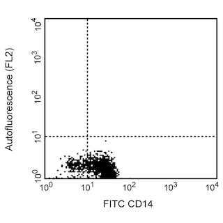
Immunofluorescent staining and flow cytometric analysis: PE Mouse Anti-Human CD119 (Cat. No. 558937) can be used for the immunofluorescent staining (≤ 1 µg antibody/10[6] cells) and flow cytometric analysis of the levels of membrane IFN-γRα expressed by human cell lines or human lymphoid cells. An appropriate immunoglobulin isotype control is PE Mouse IgG2b, κ Isotype Control (Cat. No. 555058).
Since GIR-94 is a non-neutralizing antibody, it can be used for the unobstructed immunofluorescent staining and flow cytometric analysis of cells in systems where the IFN-γ ligand is present. Based on our testing results,(data not shown) the presence of exogenous recombinant human IFN-γ at levels ≤ 50 ng/10[6] cells was insufficient to inhibit the binding of GIR-94 (at 0.06 µg mAb/1 million cells).
Product Notices
- Since applications vary, each investigator should titrate the reagent to obtain optimal results.
- An isotype control should be used at the same concentration as the antibody of interest.
- Caution: Sodium azide yields highly toxic hydrazoic acid under acidic conditions. Dilute azide compounds in running water before discarding to avoid accumulation of potentially explosive deposits in plumbing.
- For fluorochrome spectra and suitable instrument settings, please refer to our Multicolor Flow Cytometry web page at www.bdbiosciences.com/colors.
- Please refer to www.bdbiosciences.com/us/s/resources for technical protocols.
Companion Products


The GIR-94 antibody specifically recognizes the extracellular region of the alpha chain subunit (80-95 kDa glycoprotein) of the human interferon-γ receptor (IFN-γRα; aka, CD119). The functionally active-form of the human IFN-γ receptor consists of two (or more) subunits, with IFN-γRα responsible for IFN-γ binding and both the IFN-γR α and β chains required for the transduction of biologic responses. The IFN-γ receptor α chain (CD119) is expressed on the surface of most human cells (except mature erythrocytes) including monocytes, macrophages, T cells, B cells, NK cells, neutrophils, fibroblasts, epithelial cells, and endothelium. The ability of this antibody to bind to IFN-γ receptors of species other than human has not been determined. The immunogen used to generate this hybridoma was human IFN-γRα purified from human placenta. The GIR-94 is a non-neutralizing antibody.

Development References (4)
-
Bach EA, Aguet M, Schreiber RD. The IFN gamma receptor: a paradigm for cytokine receptor signaling. Annu Rev Immunol. 1997; 15:563-591. (Biology). View Reference
-
Greenlund AC, Schreiber RD, Goeddel DV, Pennica D. Interferon-gamma induces receptor dimerization in solution and on cells. J Biol Chem. 1997; 268(24):18103-18110. (Biology). View Reference
-
Kishimoto T. Tadamitsu Kishimoto .. et al., ed. Leucocyte typing VI : white cell differentiation antigens : proceedings of the sixth international workshop and conference held in Kobe, Japan, 10-14 November 1996. New York: Garland Pub.; 1997:818-821.
-
Sheehan KC, Calderon J, Schreiber RD. Generation and characterization of monoclonal antibodies specific for the human IFN-gamma receptor. J Immunol. 1988; 140(12):4231-4237. (Immunogen). View Reference
Please refer to Support Documents for Quality Certificates
Global - Refer to manufacturer's instructions for use and related User Manuals and Technical data sheets before using this products as described
Comparisons, where applicable, are made against older BD Technology, manual methods or are general performance claims. Comparisons are not made against non-BD technologies, unless otherwise noted.
For Research Use Only. Not for use in diagnostic or therapeutic procedures.