Skip to main content Skip to navigation
PE Mouse Anti-Human BCAM (CD239)
Alert icon
This SKU will be discontinuing Apr 2024. For additional support, contact your local applications specialist. Contact Us #
PE Mouse Anti-Human BCAM (CD239)
Two-color flow cytometric analysis of Human BCAM (CD239) expression on human peripheral erythrocytes. Human peripheral red blood cells were stained with BD Horizon™ BV421 Mouse Anti-Human CD235a antibody (Cat. No. 562938) and either PE Mouse IgG2a, κ Isotype Control (Cat. No. 554648; Left Plot) or PE Mouse Anti-Human BCAM (CD239) antibody (Cat. No. 566661; Right Plot) at 0.5 µg/test. Two-color flow cytometric dot plots showing the correlated expression of CD235a versus BCAM (CD239) [or Ig Isotype control staining] were derived from gated events with the forward and side light-scatter characteristics of viable red blood cells. Flow cytometric analysis was performed using a BD FACSCanto™ II Flow Cytometer System. Data shown on this Technical Data Sheet are not lot specific.
PE Mouse Anti-Human BCAM (CD239)
Immunohistochemical staining of Human BCAM (CD239) in human tonsil. Frozen sections of human tonsil were fixed with cold acetone, washed, and then stained with either Purified Mouse IgG2a κ Isotype Control (Cat. No. 550339; Left Image) or Purified Mouse Anti-Human BCAM (CD239) antibody (Right Image). A three-step staining procedure that employs a Biotin Goat Anti-Mouse Immunoglobulin (Cat. No. 550337), Streptavidin-Horseradish Peroxidase (HRP) (Cat. No.550946), and the DAB Substrate Kit (Cat. No. 550880) was used to develop the primary staining reagents. As shown in the Right Panel, the Purified Mouse Anti-Human BCAM (CD239) antibody specifically stained vascular endothelial cells of vessels. Original magnification: 20×.
Two-color flow cytometric analysis of Human BCAM (CD239) expression on human peripheral erythrocytes. Human peripheral red blood cells were stained with BD Horizon™ BV421 Mouse Anti-Human CD235a antibody (Cat. No. 562938) and either PE Mouse IgG2a, κ Isotype Control (Cat. No. 554648; Left Plot) or PE Mouse Anti-Human BCAM (CD239) antibody (Cat. No. 566661; Right Plot) at 0.5 µg/test. Two-color flow cytometric dot plots showing the correlated expression of CD235a versus BCAM (CD239) [or Ig Isotype control staining] were derived from gated events with the forward and side light-scatter characteristics of viable red blood cells. Flow cytometric analysis was performed using a BD FACSCanto™ II Flow Cytometer System. Data shown on this Technical Data Sheet are not lot specific.
Immunohistochemical staining of Human BCAM (CD239) in human tonsil. Frozen sections of human tonsil were fixed with cold acetone, washed, and then stained with either Purified Mouse IgG2a κ Isotype Control (Cat. No. 550339; Left Image) or Purified Mouse Anti-Human BCAM (CD239) antibody (Right Image). A three-step staining procedure that employs a Biotin Goat Anti-Mouse Immunoglobulin (Cat. No. 550337), Streptavidin-Horseradish Peroxidase (HRP) (Cat. No.550946), and the DAB Substrate Kit (Cat. No. 550880) was used to develop the primary staining reagents. As shown in the Right Panel, the Purified Mouse Anti-Human BCAM (CD239) antibody specifically stained vascular endothelial cells of vessels. Original magnification: 20×.
Product Details
Down Arrow Up Arrow


BD Pharmingen™
B-CAM; BCAM; Lutheran glycoprotein; MSK19
Human (QC Testing)
Mouse C57BL/6 IgG2a, κ
Human BCAM Transfected Cell Line
Flow cytometry (Routinely Tested)
0.2 mg/ml
AB_2869818
Aqueous buffered solution containing ≤0.09% sodium azide.
RUO


Preparation And Storage

Store undiluted at 4°C and protected from prolonged exposure to light. Do not freeze. The monoclonal antibody was purified from tissue culture supernatant or ascites by affinity chromatography. The antibody was conjugated with R-PE under optimum conditions, and unconjugated antibody and free PE were removed.

Product Notices

  1. Since applications vary, each investigator should titrate the reagent to obtain optimal results.
  2. An isotype control should be used at the same concentration as the antibody of interest.
  3. Caution: Sodium azide yields highly toxic hydrazoic acid under acidic conditions. Dilute azide compounds in running water before discarding to avoid accumulation of potentially explosive deposits in plumbing.
  4. Please refer to www.bdbiosciences.com/us/s/resources for technical protocols.
  5. For fluorochrome spectra and suitable instrument settings, please refer to our Multicolor Flow Cytometry web page at www.bdbiosciences.com/colors.
566661 Rev. 1
Antibody Details
Down Arrow Up Arrow
B64

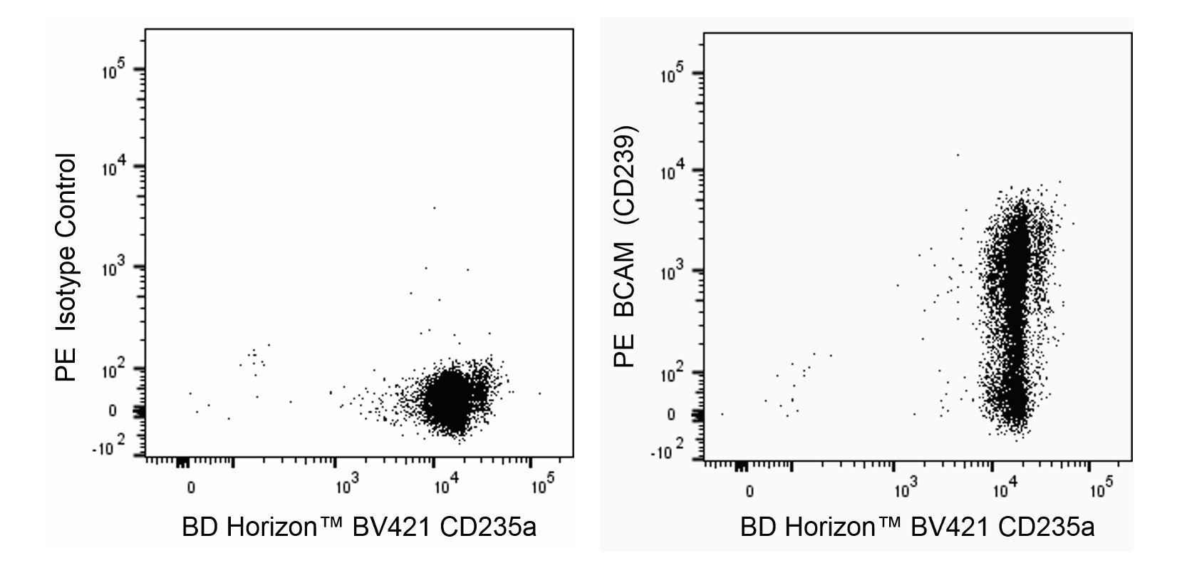
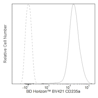
The B64 monoclonal antibody specifically recognizes Basal Cell Adhesion Molecule (BCAM, B-CAM) which is also known as CD239, Lutheran blood group antigen (Lu), or Lutheran glycoprotein. BCAM (CD239) is a type I transmembrane glycoprotein that is encoded by BCAM [basal cell adhesion molecule (Lutheran blood group)] which belongs to the Ig gene superfamily. Full-length BCAM (CD239) is comprised of an extracellular region with two N-terminal V-type Ig-like domains followed by three C2-type Ig-like domains, a transmembrane region, and cytoplasmic domain of 59 amino acid residues. The extracellular region of this adhesion molecule functions as a receptor for α5-chain containing laminins which are found in basement membranes whereas its cytoplasmic tail may have signal-transduction functions. BCAM (CD239) is expressed on erythrocytes and on the basal layer of vascular endothelial cells or some epithelial cells. BCAM (CD239) is highly expressed on sickle red cells and may play a role in vaso-occlusive crises in sickle cell disease. It is also upregulated upon malignant transformation of some cell types including those found in ovarian carcinomas.

566661 Rev. 1
Format Details
Down Arrow Up Arrow
PE
R-Phycoerythrin (PE), is part of the BD family of Phycobiliprotein dyes. This fluorochrome is a multimeric fluorescent phycobiliprotein with excitation maximum (Ex Max) of 496 nm and 566 nm and an emission maximum (Em Max) at 576 nm. PE is designed to be excited by the Blue (488 nm), Green (532 nm) and Yellow-Green (561 nm) lasers and detected using an optical filter centered near 575 nm (e.g., a 575/26-nm bandpass filter). As PE is excited by multiple lasers, this can result in cross-laser excitation and fluorescence spillover on instruments with various combinations of Blue, Green, and Yellow-Green lasers. Please ensure that your instrument’s configurations (lasers and optical filters) are appropriate for this dye.
altImg
PE
Yellow-Green 488 nm, 532 nm, 561 nm
496 nm, 566 nm
576 nm
566661 Rev.1
Citations & References
Down Arrow Up Arrow

Development References (4)

  1. Kikkawa Y, Miner JH. Review: Lutheran/B-CAM: a laminin receptor on red blood cells and in various tissues.. Connect Tissue Res. 2005; 46(4-5):193-9. (Biology). View Reference
  2. Wautier MP, El Nemer W, Gane P, et al. Increased adhesion to endothelial cells of erythrocytes from patients with polycythemia vera is mediated by laminin alpha5 chain and Lu/BCAM.. Blood. 2007; 110(3):894-901. (Biology). View Reference
  3. Zen Q, Cottman M, Truskey G, Fraser R, Telen MJ. Critical factors in basal cell adhesion molecule/lutheran-mediated adhesion to laminin.. J Biol Chem. 1999; 274(2):728-34. (Biology). View Reference
  4. Zola H, Swart B, Nicholson I, Voss E. CD239. In: Zola H. Leukocyte and stromal cell molecules : the CD markers. Hoboken, N.J.: Wiley-Liss; 2007:393-394.
View All (4) View Less
566661 Rev. 1

Please refer to Support Documents for Quality Certificates


Global - Refer to manufacturer's instructions for use and related User Manuals and Technical data sheets before using this products as described


Comparisons, where applicable, are made against older BD Technology, manual methods or are general performance claims.  Comparisons are not made against non-BD technologies, unless otherwise noted.

For Research Use Only. Not for use in diagnostic or therapeutic procedures.