Skip to main content Skip to navigation
PE-CF594 Mouse Anti-PLZF
Two-color flow cytometric analysis of PLZF expression in human peripheral blood lymphocytes. Human peripheral blood mononuclear cells were fixed and permeabilized using BD Cytofix/Cytoperm™ Fixation and Permeabilization Solution (Cat. No. 554722). The cells were subsequently washed with BD Perm/Wash™ Buffer (Cat. No. 554723) and stained with Alexa Fluor® 647 Mouse Anti-Human NCAM-1 (CD56) antibody (Cat. No. 563443) and either BD Horizon™ PE-CF594 Mouse IgG1, κ Isotype Control (Cat. No. 562292; Left Plot) or BD Horizon PE-CF594 Mouse Anti-PLZF antibody (Cat. No. 565738; Right Plot). The two-color flow cytometric contour plots showing the correlated expression of CD56 versus PLZF (or Ig Isotype control staining) were derived from gated events with the forward and side light-scatter characteristics of intact lymphocytes. Flow cytometric analyses were performed using a BD LSRFortessa™ Cell Analyzer System.
PE-CF594 Mouse Anti-PLZF
Two-color flow cytometric analysis of PLZF expression in mouse thymocytes. BALB/c mouse thymocytes were stained with APC Hamster Anti-Mouse CD3e antibody (Cat. No. 553066/561826). The cells were fixed and permeabilized using BD Cytofix/Cytoperm™ Fixation and Permeabilization Solution The cells were then washed with BD Perm/Wash™ Buffer and stained with either BD Horizon PE-CF594 Mouse IgG1, κ Isotype Control (Left Plot) or BD Horizon PE-CF594 Mouse Anti-PLZF antibody (Right Plot). The two-color flow cytometric contour plots showing the correlated expression of CD3e versus PLZF (or Ig Isotype control staining) were derived from gated events with the forward and side light- scatter characteristics of intact thymocytes. Flow cytometric analyses were performed using a BD LSRFortessa™ Cell Analyzer System.
Two-color flow cytometric analysis of PLZF expression in human peripheral blood lymphocytes. Human peripheral blood mononuclear cells were fixed and permeabilized using BD Cytofix/Cytoperm™ Fixation and Permeabilization Solution (Cat. No. 554722). The cells were subsequently washed with BD Perm/Wash™ Buffer (Cat. No. 554723) and stained with Alexa Fluor® 647 Mouse Anti-Human NCAM-1 (CD56) antibody (Cat. No. 563443) and either BD Horizon™ PE-CF594 Mouse IgG1, κ Isotype Control (Cat. No. 562292; Left Plot) or BD Horizon PE-CF594 Mouse Anti-PLZF antibody (Cat. No. 565738; Right Plot). The two-color flow cytometric contour plots showing the correlated expression of CD56 versus PLZF (or Ig Isotype control staining) were derived from gated events with the forward and side light-scatter characteristics of intact lymphocytes. Flow cytometric analyses were performed using a BD LSRFortessa™ Cell Analyzer System.
Two-color flow cytometric analysis of PLZF expression in mouse thymocytes. BALB/c mouse thymocytes were stained with APC Hamster Anti-Mouse CD3e antibody (Cat. No. 553066/561826). The cells were fixed and permeabilized using BD Cytofix/Cytoperm™ Fixation and Permeabilization Solution The cells were then washed with BD Perm/Wash™ Buffer and stained with either BD Horizon PE-CF594 Mouse IgG1, κ Isotype Control (Left Plot) or BD Horizon PE-CF594 Mouse Anti-PLZF antibody (Right Plot). The two-color flow cytometric contour plots showing the correlated expression of CD3e versus PLZF (or Ig Isotype control staining) were derived from gated events with the forward and side light- scatter characteristics of intact thymocytes. Flow cytometric analyses were performed using a BD LSRFortessa™ Cell Analyzer System.
Product Details
Down Arrow Up Arrow


BD Horizon™
Zbtb16; zinc finger and BTB domain containing 16; zinc finger protein 145
Human (QC Testing), Mouse (Tested in Development)
Mouse IgG1, κ
Mouse PLZF Recombinant Protein
Intracellular staining (flow cytometry) (Routinely Tested)
0.2 mg/ml
AB_2739339
Aqueous buffered solution containing BSA and ≤0.09% sodium azide.
RUO


Preparation And Storage

Store undiluted at 4°C and protected from prolonged exposure to light. Do not freeze. The antibody was conjugated with BD Horizon™ PE-CF594 under optimum conditions, and unconjugated antibody and free PE-CF594 were removed. The monoclonal antibody was purified from tissue culture supernatant or ascites by affinity chromatography.

Product Notices

  1. Since applications vary, each investigator should titrate the reagent to obtain optimal results.
  2. An isotype control should be used at the same concentration as the antibody of interest.
  3. When excited by the yellow-green (561-nm) laser, the fluorescence may be brighter than when excited by the blue (488-nm) laser.
  4. Please observe the following precautions: Absorption of visible light can significantly alter the energy transfer occurring in any tandem fluorochrome conjugate; therefore, we recommend that special precautions be taken (such as wrapping vials, tubes, or racks in aluminum foil) to prevent exposure of conjugated reagents, including cells stained with those reagents, to room illumination.
  5. Because of the broad absorption spectrum of the tandem fluorochrome, extra care must be taken when using multi-laser cytometers, which may directly excite both PE and CF™594.
  6. CF™ is a trademark of Biotium, Inc.
  7. This product is provided under an Agreement between BIOTIUM and BD Biosciences. The manufacture, use, sale, offer for sale, or import of this product is subject to one or more patents or pending applications owned or licensed by Biotium, Inc. This product, and only in the amount purchased by buyer, may be used solely for buyer’s own internal research, in a manner consistent with the accompanying product literature. No other right to use, sell or otherwise transfer (a) this product, or (b) its components is hereby granted expressly, by implication or by estoppel. This product is for research use only. Diagnostic uses require a separate license from Biotium, Inc. For information on purchasing a license to this product including for purposes other than research, contact Biotium, Inc., 3159 Corporate Place, Hayward, CA 94545, Tel: (510) 265-1027. Fax: (510) 265-1352. Email: btinfo@biotium.com.
  8. Texas Red is a registered trademark of Molecular Probes, Inc., Eugene, OR.
  9. Source of all serum proteins is from USDA inspected abattoirs located in the United States.
  10. Caution: Sodium azide yields highly toxic hydrazoic acid under acidic conditions. Dilute azide compounds in running water before discarding to avoid accumulation of potentially explosive deposits in plumbing.
  11. For fluorochrome spectra and suitable instrument settings, please refer to our Multicolor Flow Cytometry web page at www.bdbiosciences.com/colors.
  12. Please refer to www.bdbiosciences.com/us/s/resources for technical protocols.
565738 Rev. 1
Antibody Details
Down Arrow Up Arrow
R17-809

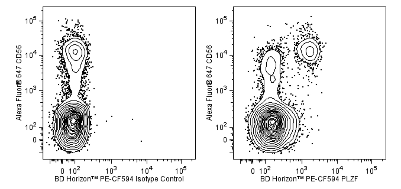
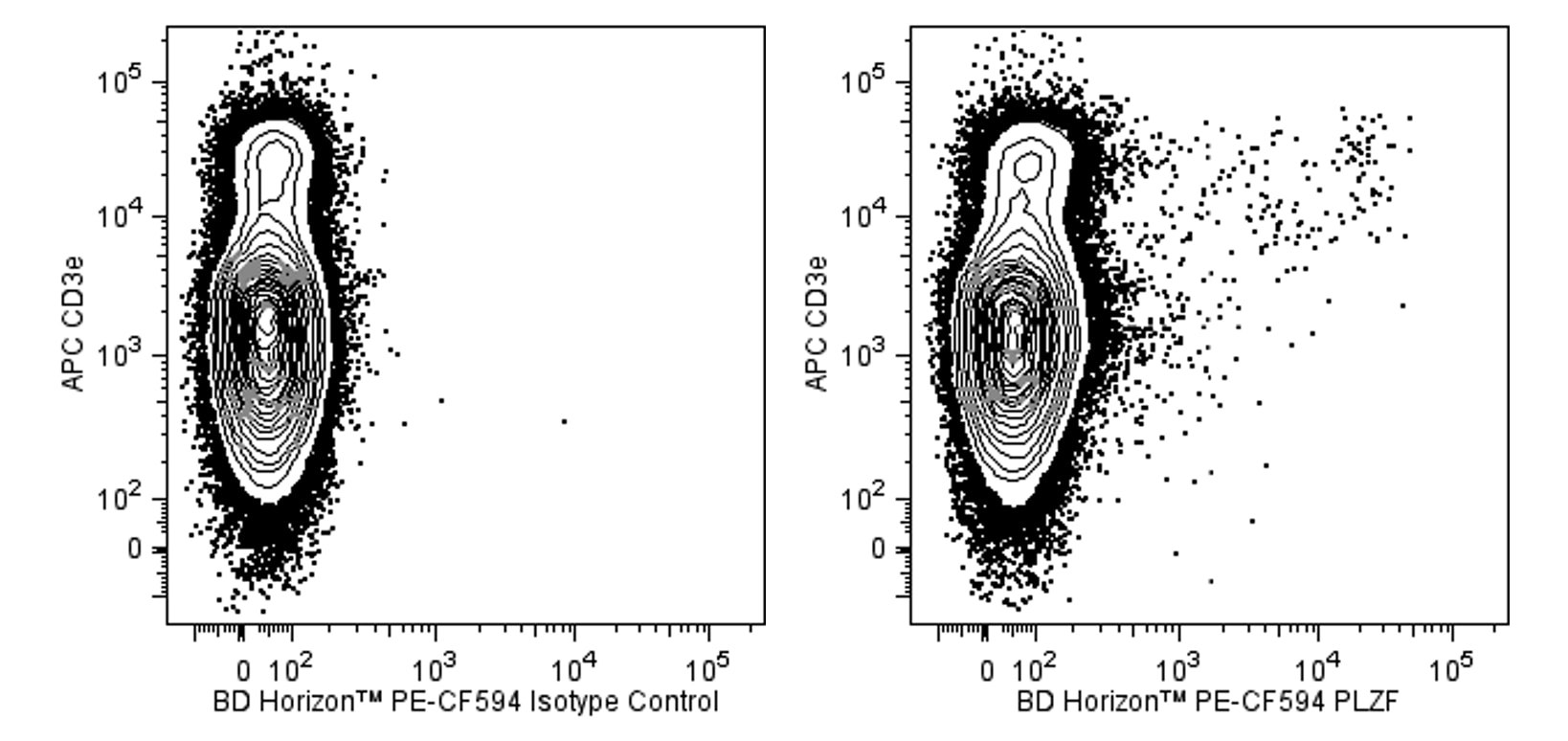
The R17-809 monoclonal antibody recognizes promyelocytic leukemia zinc finger protein (PLZF). PLZF is also known as zinc finger and BTB domain containing 16 (ZBTB16) and Zinc finger protein 145. PLZF is a member of the BTB/POZ-ZF family of transcription factors that includes Th-POK (Zbtb7b) which is involved in CD4+ T cell fate determination. PLZF serves multiple functions. In the immune system, PLZF is involved in the developmental regulation of innate immune lymphocytes including NKT cells, a subset of γδ T cells, and mucosal associated invariant T (MAIT) cells. The R17-809 hybridoma was generated from a mouse immunized with recombinant mouse PLZF protein. The R17-809 antibody crossreacts with both mouse and human PLZF.

This antibody is conjugated to BD Horizon PE-CF594, which has been developed exclusively by BD Biosciences as a better alternative to PE-Texas Red®. PE-CF594 excites and emits at similar wavelengths to PE-Texas Red® yet exhibits improved brightness and spectral characteristics. Due to PE having maximal absorption peaks at 496 nm and 564 nm, PE-CF594 can be excited by the blue (488-nm), green (532-nm) and yellow-green (561-nm) lasers and can be detected with the same filter set as PE-Texas Red® (eg 610/20-nm filter).

565738 Rev. 1
Format Details
Down Arrow Up Arrow
PE-CF594
BD Horizon™ PE-CF594 dye is a part of the BD PE family of dyes. This tandem fluorochrome is comprised of a R-Phycoerythrin (PE) donor that has excitation maxima (Ex Max) of 496-nm and 566-nm and an acceptor dye with an emission maximum (Em Max) at 615-nm. PE-CF594, driven by BD innovation, is designed to be excited by the blue (488-nm), Green (532-nm) and yellow-green (561-nm) lasers and detected using an optical filter centered near 615 nm (e.g., a 610/20-nm bandpass filter). The donor dye can be excited by the Blue (488-nm), Green (532-nm) and yellow-green (561-nm) lasers and the acceptor dye can be excited by the green (532-nm) laser resulting in cross-laser excitation and fluorescence spillover. Please ensure that your instrument’s configurations (lasers and optical filters) are appropriate for this dye.
altImg
PE-CF594
Yellow-Green 488 nm, 532 nm, 561 nm
496 nm, 566 nm
615 nm
565738 Rev.1
Citations & References
Down Arrow Up Arrow

Development References (4)

  1. Alonzo ES, Gottschalk RA, Das J, et al. Development of promyelocytic zinc finger and ThPOK-expressing innate gamma delta T cells is controlled by strength of TCR signaling and Id3. J Immunol. 2010; 184(3):1268-1279. (Biology). View Reference
  2. Alonzo ES, Sant'Angelo DB. Development of PLZF-expressing innate T cells. Curr Opin Immunol. 2011; 23(2):220-227. (Biology). View Reference
  3. Moon H, Park C, Lee JG, et al. Early Development in the Peritoneal Cavity of CD49dhigh Th1 Memory Phenotype CD4+ T Cells with Enhanced B Cell Helper Activity.. J Immunol. 2015; 195(2):564-75. (Clone-specific: Flow cytometry). View Reference
  4. Savage AK, Constantinides MG, Han J, et al. The transcription factor PLZF directs the effector program of the NKT cell lineage. Immunity. 2008; 29(3):391-403. (Biology). View Reference
View All (4) View Less
565738 Rev. 1

Please refer to Support Documents for Quality Certificates


Global - Refer to manufacturer's instructions for use and related User Manuals and Technical data sheets before using this products as described


Comparisons, where applicable, are made against older BD Technology, manual methods or are general performance claims.  Comparisons are not made against non-BD technologies, unless otherwise noted.

For Research Use Only. Not for use in diagnostic or therapeutic procedures.