
Preparation And Storage
Product Notices
- Since applications vary, each investigator should titrate the reagent to obtain optimal results.
- Please refer to www.bdbiosciences.com/us/s/resources for technical protocols.
- The Alexa Fluor®, Pacific Blue™, and Cascade Blue® dye antibody conjugates in this product are sold under license from Molecular Probes, Inc. for research use only, excluding use in combination with microarrays, or as analyte specific reagents. The Alexa Fluor® dyes (except for Alexa Fluor® 430), Pacific Blue™ dye, and Cascade Blue® dye are covered by pending and issued patents.
- Alexa Fluor® 647 fluorochrome emission is collected at the same instrument settings as for allophycocyanin (APC).
- Alexa Fluor® is a registered trademark of Molecular Probes, Inc., Eugene, OR.
- Caution: Sodium azide yields highly toxic hydrazoic acid under acidic conditions. Dilute azide compounds in running water before discarding to avoid accumulation of potentially explosive deposits in plumbing.
- For fluorochrome spectra and suitable instrument settings, please refer to our Multicolor Flow Cytometry web page at www.bdbiosciences.com/colors.
- An isotype control should be used at the same concentration as the antibody of interest.
Companion Products


.png?imwidth=320)
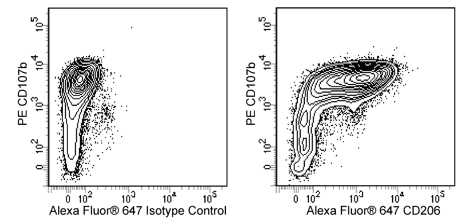
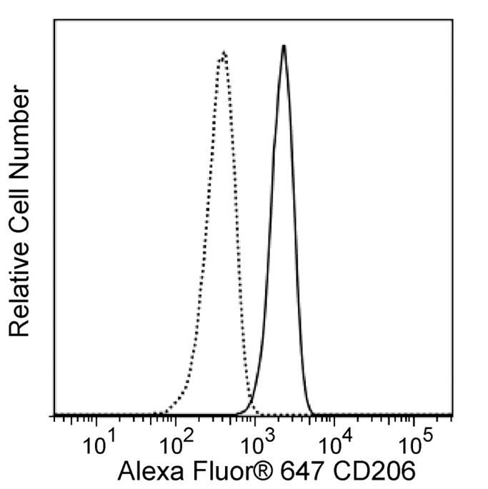
The MR5D3 monoclonal antibody specifically binds to CD206 which is also known as the Macrophage mannose receptor (MMR, MR) or Mannose receptor, C type 1 (Mrc1). CD206 is a type I transmembrane glycoprotein of approximately 175 kDa that belongs to the C-type lectin superfamily. It is expressed at the cell surface and intracellularly by macrophages, Langerhans cells, dendritic cells, and endothelial cells within hepatic and lymphoid tissues. This pattern recognition receptor binds to endogenous and microbial glycoconjugates containing mannose, fucose, or N-acetylglucosamine through its C-type lectin-like carbohydrate recognition domains (CRD). CD206 also contains a cysteine-rich domain that enables binding to sulfated carbohydrate antigens. This receptor enables macrophages and other specialized cells to maintain tissue homeostasis as well as to internalize microbes or their components by phagocytosis or endocytosis. CD206 thus plays important roles in mediating innate immunity, e.g., enabling phagocytosis, as well as in processing and presenting antigens for the generation and expression of adaptive immunity. Moreover, CD206 has been associated with leucocyte homing and cancer cell metastasis.
Development References (5)
-
Akbarshahi H, Menzel M, Posaric Bauden M, Rosendahl A, Andersson R. Enrichment of murine CD68+ CCR2+ and CD68+ CD206+ lung macrophages in acute pancreatitis-associated acute lung injury. PLoS ONE. 2012; 7(10):e42654. (Biology). View Reference
-
Burgdorf S, Lukacs-Kornek V, Kurts C. The mannose receptor mediates uptake of soluble but not of cell-associated antigen for cross-presentation. J Immunol. 2006; 176(11):6770-6776. (Biology). View Reference
-
Marttila-Ichihara F, Turja R, Miiluniemi M, et al. Macrophage mannose receptor on lymphatics controls cell trafficking. Blood. 2008; 112(1):64-72. (Clone-specific: Immunofluorescence, Immunohistochemistry). View Reference
-
McKenzie EJ, Taylor PR, Stillion RJ, et al. J Immunol. 2007; 178(8):4975-4983. (Clone-specific: Flow cytometry). View Reference
-
Zamze S, Martinez-Pomares L, Jones H, et al. Recognition of bacterial capsular polysaccharides and lipopolysaccharides by the macrophage mannose receptor. J Biol Chem. 2002; 277(44):41613-41623. (Immunogen: Dot Blot, ELISA, Flow cytometry, Immunoaffinity chromatography, Immunohistochemistry, Immunoprecipitation). View Reference
Please refer to Support Documents for Quality Certificates
Global - Refer to manufacturer's instructions for use and related User Manuals and Technical data sheets before using this products as described
Comparisons, where applicable, are made against older BD Technology, manual methods or are general performance claims. Comparisons are not made against non-BD technologies, unless otherwise noted.
For Research Use Only. Not for use in diagnostic or therapeutic procedures.