-
Your selected country is
Middle East / Africa
- Change country/language
Old Browser
This page has been recently translated and is available in French now.
Looks like you're visiting us from {countryName}.
Would you like to stay on the current country site or be switched to your country?


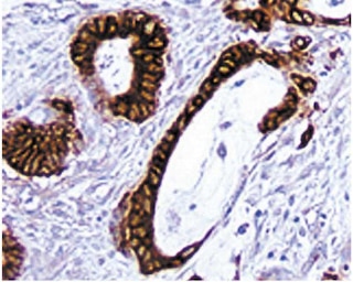
Immunohistochemical analysis of clusterin expression in human tonsil. Formalin-fixed, paraffin-embedded sections of human tonsil were stained Purified Mouse Anti-human Clusterin (Cat. No. 552886), followed by Biotin Goat Anti-Mouse Ig (Multiple Adsorption)(Cat. No. 550337) and Streptavidin HRP (Cat. No. 550946). Dendritic cells in the follicle and basement membrane of epithelial cells and fibrocytes expressed clusterin. Magnification 20X.


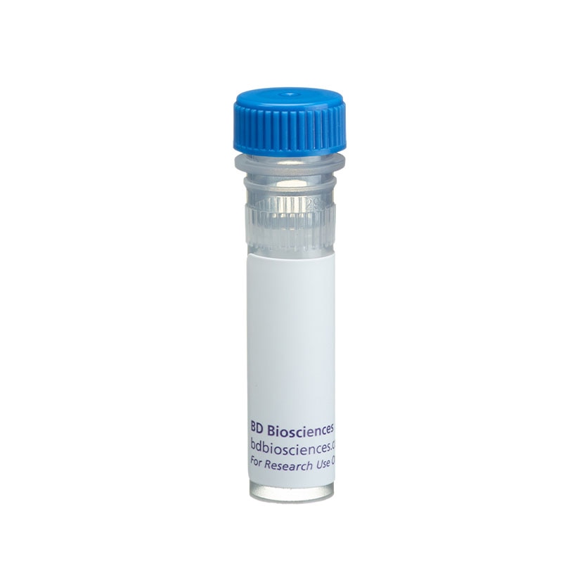
BD Pharmingen™ Purified Mouse Anti-human Clusterin

Regulatory Status Legend
Any use of products other than the permitted use without the express written authorization of Becton, Dickinson and Company is strictly prohibited.
Preparation And Storage
Recommended Assay Procedures
Immunohistochemistry: Purified Mouse Anti-human Clusterin was tested for immunohistochemical staining of acetone-fixed frozen sections and zinc-fixed and formalin-fixed, paraffin-embedded sections. The tissue tested was human tonsil. The antibody stains dendritic cells in the follicle and basement membrane of epithelial cells. The isotype control recommended for use with this antibody is Purified Mouse IgG1 κ Isotype Control (Cat. No. 550878). For optimal indirect immunohistochemical staining, the E5 antibody should be titrated (1:10 to 1:50 dilution) and visualized via a three-step staining procedure in combination with Biotin Goat Anti-Mouse Ig (Multiple Adsorption)(Cat. No. 550337) as the secondary antibody and Streptavidin-HRP (Cat. No. 550946) together with the DAB Detection System (Cat. No. 550880). More conveniently, the Anti-Mouse Ig HRP Detection Kit (Cat. No. 551011), which contains Biotin Goat Anti-Mouse Ig (Multiple Adsorption), antibody diluent, Streptavidin-HRP and DAB substrate, can be used for staining. A detailed protocol of the immunohistochemical procedure is available at http://www.bdbiosciences.com/us/s/resources.
Product Notices
- Since applications vary, each investigator should titrate the reagent to obtain optimal results.
- An isotype control should be used at the same concentration as the antibody of interest.
- Caution: Sodium azide yields highly toxic hydrazoic acid under acidic conditions. Dilute azide compounds in running water before discarding to avoid accumulation of potentially explosive deposits in plumbing.
- Source of all serum proteins is from USDA inspected abattoirs located in the United States.
- Please refer to http://regdocs.bd.com to access safety data sheets (SDS).
- Sodium azide is a reversible inhibitor of oxidative metabolism; therefore, antibody preparations containing this preservative agent must not be used in cell cultures nor injected into animals. Sodium azide may be removed by washing stained cells or plate-bound antibody or dialyzing soluble antibody in sodium azide-free buffer. Since endotoxin may also affect the results of functional studies, we recommend the NA/LE (No Azide/Low Endotoxin) antibody format, if available, for in vitro and in vivo use.
- Please refer to www.bdbiosciences.com/us/s/resources for technical protocols.
Companion Products


The E5 antibody reacts with human clusterin. Clusterin was originally isolated and characterized from glomerular immune deposits of patients with renal glomerulonephritis. Clusterin, an 80 kD disulfide-linked, heterodimeric glycoprotein (previously named SP-40,40 [serum protein 40 kD,40kD]) was shown to be part of the SC5b-9 complex of complement, yet separate and distinct from the S protein. Clusterins have since been identified in a number of mammalian species and have been implicated in diverse cellular activities, however, the exact role remains to be elucidated.
Development References (3)
-
Murphy BF, Kirszbaum L, Walker ID, d'Apice AJ. SP-40,40, a newly identified normal human serum protein found in the SC5b-9 complex of complement and in the immune deposits in glomerulonephritis. J Clin Invest. 1988; 81(6):1858-1864. (Clone-specific: Western blot). View Reference
-
Rosenberg ME, Silkensen J. Clusterin: physiologic and pathophysiologic considerations. Int J Biochem Cell Biol. 1995; 27(7):633-645. (Clone-specific). View Reference
-
Wellmann A, Thieblemont C, Pittaluga S, et al. Detection of differentially expressed genes in lymphomas using cDNA arrays: identification of clusterin as a new diagnostic marker for anaplastic large-cell lymphomas. Blood. 2000; 96(2):398-404. (Clone-specific: Western blot). View Reference
Please refer to Support Documents for Quality Certificates
Global - Refer to manufacturer's instructions for use and related User Manuals and Technical data sheets before using this products as described
Comparisons, where applicable, are made against older BD Technology, manual methods or are general performance claims. Comparisons are not made against non-BD technologies, unless otherwise noted.
For Research Use Only. Not for use in diagnostic or therapeutic procedures.
Report a Site Issue
This form is intended to help us improve our website experience. For other support, please visit our Contact Us page.