

Preparation And Storage
Recommended Assay Procedures
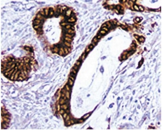
Immunohistochemistry: The M17/4 antibody specific for mouse CD11a is recommended to test for immunohistochemical staining of acetone-fixed frozen sections. Tissues tested were mouse spleen and thymus. The antibody stains all leukocytes. The isotype control recommended for use with this antibody is purified rat IgG2a (Cat. No. 559073). For optimal indirect immunohistochemical staining, the M17/4 antibody should be titrated (1:10 to 1:50 dilution) and visualized via a three-step staining procedure in combination with polyclonal, biotin conjugated anti-rat Igs (multiple adsorbed) (Cat. No. 559286) as the secondary antibody and Streptavidin-HRP (Cat. No. 550946) together with the DAB detection system (Cat. No. 550880). A detailed protocol of the immunohistochemical procedure can be found on our website www.bdbiosciences.com/support/resources. The clone M17/4 is not recommended for formalin-fixed paraffin embedded sections.
Product Notices
- Since applications vary, each investigator should titrate the reagent to obtain optimal results.
- Caution: Sodium azide yields highly toxic hydrazoic acid under acidic conditions. Dilute azide compounds in running water before discarding to avoid accumulation of potentially explosive deposits in plumbing.
- Source of all serum proteins is from USDA inspected abattoirs located in the United States.
- An isotype control should be used at the same concentration as the antibody of interest.
- This antibody has been developed for the immunohistochemistry application. However, a routine immunohistochemistry test is not performed on every lot. Researchers are encouraged to titrate the reagent for optimal performance.
- Please refer to www.bdbiosciences.com/us/s/resources for technical protocols.
Companion Products


The M17/4 antibody reacts with the 180 kDa αL chain of LFA-1 (CD11a/CD18, αLβ2 integrin), a heterodimeric surface glycoprotein expressed on almost all leukocytes. CD8a+CD8b- intestinal intraepithelial T lymphocytes, which are believed to be thymus independent, do not express CD11a. LFA-1 mediates a variety of heterotypic and homotypic intercellular adhesions through interaction with ICAM-1 (CD54) and ICAM-2 (CD102), including participation in the immunological synapses between CD8+ T lymphocytes and antigen-presenting cells. mAb M17/4 blocks a variety of LFA-1-mediated cells interactions in vitro, and costimulatory effects have also been described. In vivo treatment with M17/4 mAb reduces the severity of graft-versus-host reactions, prolongs allograft survival, inhibits the development of autoimmunity, and blocks substance P-induced leukocyte migration. The M17/4 and 2D7 (Cat. No. 553120) antibodies are reported to recognize different epitopes of the CD11a molecule.
Development References (10)
-
Driessens MH, van Hulten P, Zuurbier A, La Riviere G, Roos E. Inhibition and stimulation of LFA-1 and Mac-1 functions by antibodies against murine CD18. Evidence that the LFA-1 binding sites for ICAM-1, -2, and -3 are distinct. J Leukoc Biol. 1996; 60(6):758-765. (Clone-specific: Immunohistochemistry). View Reference
-
Harning R, Pelletier J, Lubbe K, Takei F, Merluzzi VJ. Reduction in the severity of graft-versus-host disease and increased survival in allogenic mice by treatment with monoclonal antibodies to cell adhesion antigens LFA-1 alpha and MALA-2. Transplantation. 1991; 52(5):842-845. (Clone-specific: Immunohistochemistry). View Reference
-
Kuhlman P, Moy VT, Lollo BA, Brian AA. The accessory function of murine intercellular adhesion molecule-1 in T lymphocyte activation. Contributions of adhesion and co-activation. J Immunol. 1991; 146(6):1773-1782. (Clone-specific: (Co)-stimulation). View Reference
-
Larson RS, Springer TA. Structure and function of leukocyte integrins. Immunol Rev. 1990; 114:181-217. (Biology). View Reference
-
Sanchez-Madrid F, Davignon D, Martz E, Springer TA. Antigens involved in mouse cytolytic T-lymphocyte (CTL)-mediated killing: functional screening and topographic relationship. Cell Immunol. 1982; 73(1):1-11. (Immunogen: Immunohistochemistry). View Reference
-
Sanchez-Madrid F, Simon P, Thompson S, Springer TA. Mapping of antigenic and functional epitopes on the alpha- and beta-subunits of two related mouse glycoproteins involved in cell interactions, LFA-1 and Mac-1. J Exp Med. 1983; 158(2):586-602. (Clone-specific: Immunohistochemistry). View Reference
-
Sanders VM, Vitetta ES. B cell-associated LFA-1 and T cell-associated ICAM-1 transiently cluster in the area of contact between interacting cells. Cell Immunol. 1991; 132(1):45-55. (Clone-specific: Immunohistochemistry). View Reference
-
Springer TA, Davignon D, Ho MK, Kurzinger K, Martz E, Sanchez-Madrid F. LFA-1 and Lyt-2,3, molecules associated with T lymphocyte-mediated killing; and Mac-1, an LFA-1 homologue associated with complement receptor function. Immunol Rev. 1982; 68:171-195. (Clone-specific: Immunohistochemistry). View Reference
-
Springer TA. Traffic signals for lymphocyte recirculation and leukocyte emigration: the multistep paradigm. Cell. 1994; 76(2):301-314. (Biology). View Reference
-
Zhao Y, Iwata M. Cross-linking of the TCR-CD3 complex with CD4, CD8 or LFA-1 induces an anti-apoptotic signal in thymocytes: the signal is canceled by FK506. Int Immunol. 1995; 7(9):1387-1396. (Clone-specific: (Co)-stimulation). View Reference
Please refer to Support Documents for Quality Certificates
Global - Refer to manufacturer's instructions for use and related User Manuals and Technical data sheets before using this products as described
Comparisons, where applicable, are made against older BD Technology, manual methods or are general performance claims. Comparisons are not made against non-BD technologies, unless otherwise noted.
For Research Use Only. Not for use in diagnostic or therapeutic procedures.