
Preparation And Storage
Recommended Assay Procedures
BD® CompBeads can be used as surrogates to assess fluorescence spillover (compensation). When fluorochrome conjugated antibodies are bound to BD® CompBeads, they have spectral properties very similar to cells. However, for some fluorochromes there can be small differences in spectral emissions compared to cells, resulting in spillover values that differ when compared to biological controls. It is strongly recommended that when using a reagent for the first time, users compare the spillover on cell and BD® CompBeads to ensure that BD® CompBeads are appropriate for your specific cellular application.
Product Notices
- Please refer to www.bdbiosciences.com/us/s/resources for technical protocols.
- This reagent has been pre-diluted for use at the recommended Volume per Test. We typically use 1 × 10^6 cells in a 100-µl experimental sample (a test).
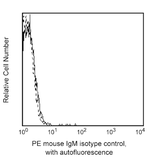
- An isotype control should be used at the same concentration as the antibody of interest.
- Caution: Sodium azide yields highly toxic hydrazoic acid under acidic conditions. Dilute azide compounds in running water before discarding to avoid accumulation of potentially explosive deposits in plumbing.
- For fluorochrome spectra and suitable instrument settings, please refer to our Multicolor Flow Cytometry web page at www.bdbiosciences.com/colors.
- Source of all serum proteins is from USDA inspected abattoirs located in the United States.
- Please refer to http://regdocs.bd.com to access safety data sheets (SDS).
- For U.S. patents that may apply, see bd.com/patents.
Companion Products


The VIM8 monoclonal antibody specifically binds to the asialylated form of human CD65. CD65 is also known as, Ceramide dodecasacharide 4c. CD65 is expressed on myeloid cells during development. Peripheral blood granulocytes express high levels of CD65when compared with monocytes that express lower levels. CD65 reportedly can serve as a ligand for CD62E (E-Selectin). CD65 has been implicated as a crucial adhesive ligand involved in the extravascular infiltration of acute myeloid leukemic (AML) cells.

Development References (7)
-
Easton EW, Schiphorst WE, van Drunen E, van der Schoot CE, van den Eijnden DH. Human myeloid alpha 3-fucosyltransferase is involved in the expression of the sialyl-Lewis(x) determinant, a ligand for E- and P-selectin. Blood. 1993; 81(11):2978-2986. (Clone-specific: Flow cytometry). View Reference
-
Gooi HC, Hounsell EF, Edwards A, Majdic O, Knapp W, Feizi T. Differences in the fine specificities of monoclonal (Class A) antibodies to human myeloid cells.. Clin Exp Immunol. 1985; 60(1):151-8. (Clone-specific: Immunofluorescence, Radioimmunoassay). View Reference
-
Knapp W., Majdic O, Blanchard D, et al. CDw65 cluster workshop report. In: Schlossman SF. Stuart F. Schlossman .. et al., ed. Leucocyte typing V : white cell differentiation antigens : proceedings of the fifth international workshop and conference held in Boston, USA, 3-7 November, 1993. Oxford: Oxford University Press; 1995:876-882.
-
Kniep B, Peter-Katalinic J, Müthing J, Majdic O, Pickl WF, Knapp W. The CDw65 monoclonal antibodies VIM-8 and VIM-11 bind to the neutral glycolipid V3FucnLc8Cer.. J Biochem. 1996; 119(3):456-62. (Clone-specific: Flow cytometry, Western blot). View Reference
-
Noguchi M, Sato N, Sugimori H, Mori K, Oshimi K. A minor E-selectin ligand, CD65, is critical for extravascular infiltration of acute myeloid leukemia cells.. Leuk Res. 2001; 25(10):847-53. (Biology). View Reference
-
Paietta E, Neuberg D, Bennett JM, et al. Low expression of the myeloid differentiation antigen CD65s, a feature of poorly differentiated AML in older adults: study of 711 patients enrolled in ECOG trials.. Leukemia. 2003; 17(8):1544-50. (Biology). View Reference
-
Stockinger H. Cluster report: CDw65. In: Knapp W. W. Knapp .. et al., ed. Leucocyte typing IV : white cell differentiation antigens. Oxford New York: Oxford University Press; 1989:836-838.
Please refer to Support Documents for Quality Certificates
Global - Refer to manufacturer's instructions for use and related User Manuals and Technical data sheets before using this products as described
Comparisons, where applicable, are made against older BD Technology, manual methods or are general performance claims. Comparisons are not made against non-BD technologies, unless otherwise noted.
For Research Use Only. Not for use in diagnostic or therapeutic procedures.