
Preparation And Storage
Product Notices
- This reagent has been pre-diluted for use at the recommended Volume per Test. We typically use 1 × 10^6 cells in a 100-µl experimental sample (a test).
- Source of all serum proteins is from USDA inspected abattoirs located in the United States.
- An isotype control should be used at the same concentration as the antibody of interest.
- Please refer to www.bdbiosciences.com/us/s/resources for technical protocols.
- Caution: Sodium azide yields highly toxic hydrazoic acid under acidic conditions. Dilute azide compounds in running water before discarding to avoid accumulation of potentially explosive deposits in plumbing.
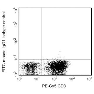
- For fluorochrome spectra and suitable instrument settings, please refer to our Multicolor Flow Cytometry web page at www.bdbiosciences.com/colors.
Companion Products


The GHI/61 monoclonal antibody specifically binds to human CD163. CD163 is also known as Scavenger receptor cysteine-rich type 1 protein M130 (M130), Hemoglobin scavenger receptor and Macrophage-associated antigen. CD163 is a 110-130 kDa transmembrane glycoprotein. CD163 is a monocyte/macrophage-restricted antigen expressed on the majority of tissue macrophages and peripheral blood monocytes. CD163 belongs to the scavenger receptor superfamily. Its expression on monocytes is upregulated upon cellular activation. CD163 expression reportedly changes on monocytes and macrophages as these cells differentiate. This finding suggests a role for this molecule in the differentiation and/or regulation of monocyte and macrophage function. CD163 may play a role in the clearance and endocytosis of hemoglobin and haptoglobin complexes by macrophages.
It has been reported (Maniecki et al., 2011) that the presence of calcium impacts the binding affinity of clone GHI/61 to CD163. There is a variation in detecting CD163 positive monocytes when the cells are prepared with different anticoagulants, where heparin was observed to have the highest inhibitory effect on clone GHI/61.

Development References (4)
-
Kishimoto T. Tadamitsu Kishimoto .. et al., ed. Leucocyte typing VI : white cell differentiation antigens : proceedings of the sixth international workshop and conference held in Kobe, Japan, 10-14 November 1996. New York: Garland Pub.; 1997.
-
Law SK, Micklem KJ, Shaw JM. A new macrophage differentiation antigen which is a member of the scavenger receptor superfamily. Eur J Immunol. 1993; 23(9):2320-2325. (Biology). View Reference
-
Maniecki MB, Etzerodt A, Moestrup S, Møller J, Graversen J. Comparative assessment of the recognition of domain-specific CD163 monoclonal antibodies in human monocytes explains wide discrepancy in reported levels of cellular surface CD163 expression. Immunobiology. 2011; 216(8):882-890. (Clone-specific: Flow cytometry, Western blot). View Reference
-
Pulford K, Micklem K, McCarthy S, Cordell J, Jones M, Mason DY. A monocyte/macrophage antigen recognized by the four antibodies GHI/61, Ber-MAC3, Ki-M8 and SM4. Immunology. 1992; 75(4):588-595. (Immunogen: Blocking, Flow cytometry, Immunoaffinity chromatography, Immunohistochemistry, Immunoprecipitation). View Reference
Please refer to Support Documents for Quality Certificates
Global - Refer to manufacturer's instructions for use and related User Manuals and Technical data sheets before using this products as described
Comparisons, where applicable, are made against older BD Technology, manual methods or are general performance claims. Comparisons are not made against non-BD technologies, unless otherwise noted.
For Research Use Only. Not for use in diagnostic or therapeutic procedures.