-
Your selected country is
Middle East / Africa
- Change country/language
Old Browser
This page has been recently translated and is available in French now.
Looks like you're visiting us from {countryName}.
Would you like to stay on the current country site or be switched to your country?
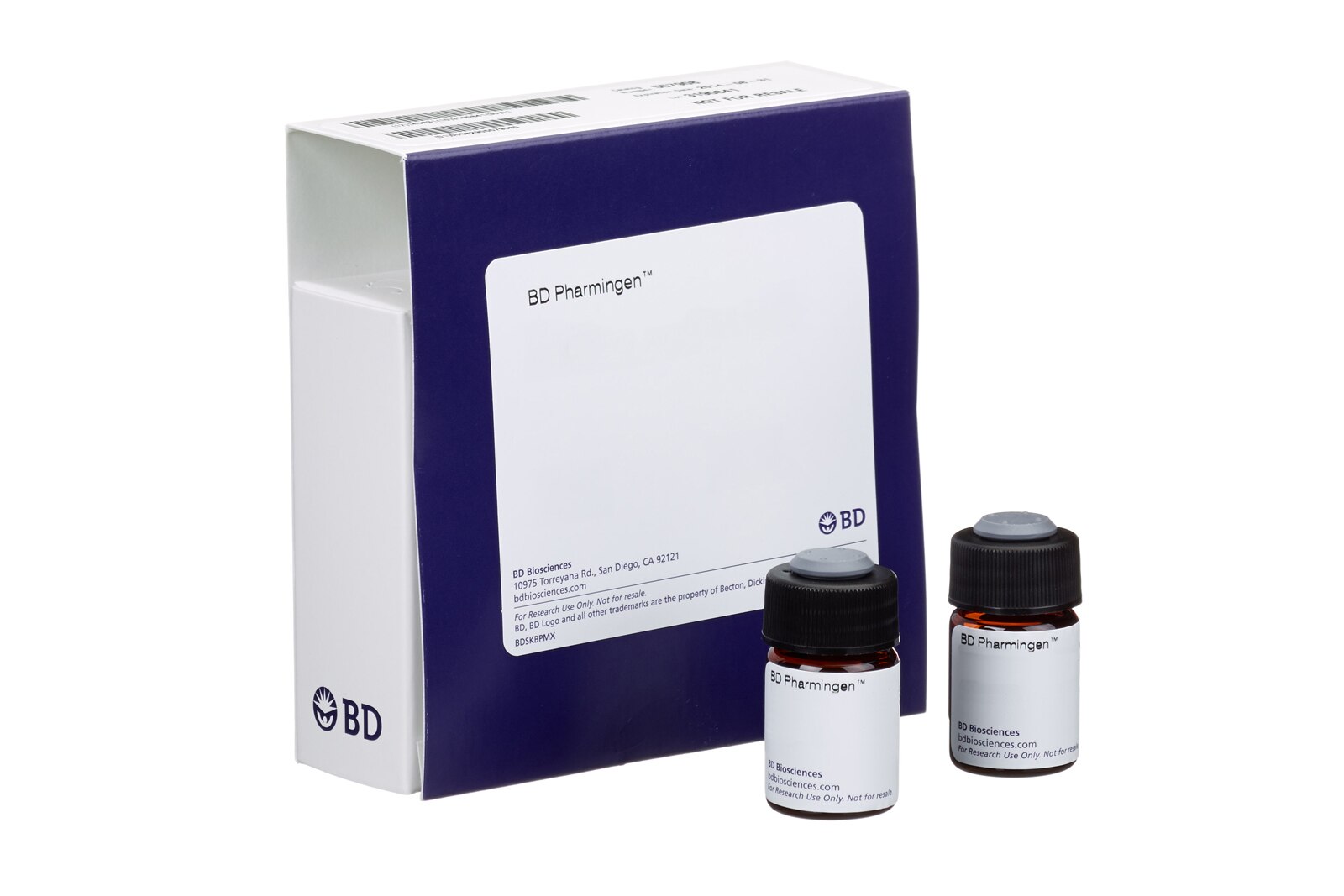
BD Pharmingen™ PE Hamster Anti-Human Bcl-2 Set
(RUO)


Profile of permeabilized peripheral blood lymphocytes analyzed on a BD FACScan™ flow cytometry instrument. Cells were stained with the PE hamster anti-human Bcl-2 antibody (shaded) or with a PE hamster IgG2 isotype control (unshaded).


BD Pharmingen™ PE Hamster Anti-Human Bcl-2 Set

PE Hamster Anti-Human Bcl-2 Set
Regulatory Status Legend
Any use of products other than the permitted use without the express written authorization of Becton, Dickinson and Company is strictly prohibited.
Description
Programmed cell death (apoptosis) is a normal physiologic process which occurs during embryonic development as well as in maintenance of tissue homeostasis. The apoptotic program is characterized by certain morphological features. These include changes in the plasma membrane such as loss of membrane asymmetry and attachment, a condensation of the cytoplasm and nucleus, and internucleosomal cleavage of DNA. In the final stages, the dying cells become fragmented into "apoptotic bodies" which are rapidly eliminated by phagocytic cells without eliciting significant inflammatory damage to surrounding cells. Members of the Bcl-2 family play a major role in regulating the cellular response to apoptotic signals. Bcl-2 is considered to be a novel proto-oncogene because it blocks apoptosis in many cell types. Bcl-2 is thought to provide selective survival advantage for cells by blocking apoptosis and thus may contribute to tumorigenesis. Bcl-2 is a 26 kDa intracellular, integral membrane protein found primarily in the nuclear envelope, endoplasmic reticulum and outer mitochondrial membrane.
Preparation And Storage
Product Notices
- This reagent has been pre-diluted for use at the recommended Volume per Test. We typically use 1 × 10^6 cells in a 100-µl experimental sample (a test).
- Please refer to www.bdbiosciences.com/us/s/resources for technical protocols.
- Although hamster immunoglobulin isotypes have not been well defined, BD Biosciences Pharmingen has grouped Armenian and Syrian hamster IgG monoclonal antibodies according to their reactivity with a panel of mouse anti-hamster IgG mAbs. A table of the hamster IgG groups, Reactivity of Mouse Anti-Hamster Ig mAbs, may be viewed at http://www.bdbiosciences.com/documents/hamster_chart_11x17.pdf.
- Caution: Sodium azide yields highly toxic hydrazoic acid under acidic conditions. Dilute azide compounds in running water before discarding to avoid accumulation of potentially explosive deposits in plumbing.
- Source of all serum proteins is from USDA inspected abattoirs located in the United States.
| Description | Quantity/Size | Part Number | EntrezGene ID |
|---|---|---|---|
| PE Hamster Anti-Human Bcl-2 | 100 Tests (1 ea) | 51-15135X | N/A |
| PE Hamster Isotype Control | 100 Tests (1 ea) | 51-66985X | N/A |
Development References (8)
-
Hockenbery D, Nuñez G, Milliman C, Schreiber RD, Korsmeyer SJ. Bcl-2 is an inner mitochondrial membrane protein that blocks programmed cell death. Nature. 1990; 348(6299):334-336. (Immunogen: Immunofluorescence, Western blot). View Reference
-
Krajewski S, Tanaka S, Takayama S, Schibler MJ, Fenton W, Reed JC. Investigation of the subcellular distribution of the bcl-2 oncoprotein: residence in the nuclear envelope, endoplasmic reticulum, and outer mitochondrial membranes. Cancer Res. 1993; 53(19):4701-4714. (Biology). View Reference
-
Martin JM, Veis D, Korsmeyer SJ, Sugden B. Latent membrane protein of Epstein-Barr virus induces cellular phenotypes independently of expression of Bcl-2. J Virol. 1993; 67(6):5269-5278. (Biology). View Reference
-
McDonnell TJ, Nunez G, Platt FM, et al. Deregulated Bcl-2-immunoglobulin transgene expands a resting but responsive immunoglobulin M and D-expressing B-cell population. Mol Cell Biol. 1990; 10(5):1901-1907. (Biology: Western blot). View Reference
-
Reed JC, Meister L, Tanaka S, et al. Differential expression of bcl2 protooncogene in neuroblastoma and other human tumor cell lines of neural origin. Cancer Res. 1991; 51(24):6529-6538. (Biology). View Reference
-
Veis DJ, Sentman CL, Bach EA, Korsmeyer SJ. Expression of the Bcl-2 protein in murine and human thymocytes and in peripheral T lymphocytes. J Immunol. 1993; 151(5):2546-2554. (Biology). View Reference
-
Williams GT. Programmed cell death: apoptosis and oncogenesis. Cell. 1991; 65(7):1097-1098. (Biology). View Reference
-
Yang J, Liu X, Bhalla K, et al. Prevention of apoptosis by Bcl-2: release of cytochrome c from mitochondria blocked. Science. 1997; 275(5303):1129-1132. (Biology). View Reference
Please refer to Support Documents for Quality Certificates
Global - Refer to manufacturer's instructions for use and related User Manuals and Technical data sheets before using this products as described
Comparisons, where applicable, are made against older BD Technology, manual methods or are general performance claims. Comparisons are not made against non-BD technologies, unless otherwise noted.
For Research Use Only. Not for use in diagnostic or therapeutic procedures.
Report a Site Issue
This form is intended to help us improve our website experience. For other support, please visit our Contact Us page.