Skip to main content Skip to navigation
PE Mouse Anti-Human CD25
Two-color flow cytometric analysis of CD25 expression on unstimulated peripheral blood lymphocytes. Human whole blood was stained with Alexa Fluor® 647 Mouse Anti-Human CD4 antibody (Cat. No. 557707) and either PE Mouse IgG1, κ Isotype Control (Cat. No. 554680; Left Plot) or PE Mouse Anti-Human CD25 antibody (Cat. No. 567214; Right Plot) at 0.25 µg/test. Erythrocytes were lysed with BD Pharm Lyse™ Lysing Buffer (Cat. No. 555899). The bivariate pseudocolor density plot showing the correlated expression of CD25 (or Ig Isotype control staining) versus CD4 was derived from gated events with the forward and side light-scatter characteristics of viable lymphocytes. Flow cytometry and data analysis were performed using a BD LSRFortessa™ X-20 Cell Analyzer System and FlowJo™ software. Data shown on this Technical Data Sheet are not lot specific.
PE Mouse Anti-Human CD25
Flow cytometric analysis of CD25 expression on activated human peripheral blood lymphocytes. Peripheral blood mononuclear cells were stimulated for 3 days with Phytohemagglutinin (PHA). The cells were stained with either PE Mouse IgG1, κ Isotype Control (dashed line histogram) or PE Mouse Anti-Human CD25 antibody (solid line histogram) at 0.25 µg/test. DAPI (4',6-Diamidino-2-Phenylindole, Dihydrochloride) Solution (Cat. No. 564907) was added to cells right before analysis. The fluorescence histogram showing CD25 expression (or Ig Isotype control staining) was derived from gated events with the forward and side light-scatter characteristics of viable (DAPI-) lymphoblasts. Flow cytometry and data analysis were performed using a BD LSRFortessa™ X-20 Cell Analyzer System and FlowJo™ software. Data shown on this Technical Data Sheet are not lot specific.
Two-color flow cytometric analysis of CD25 expression on unstimulated peripheral blood lymphocytes. Human whole blood was stained with Alexa Fluor® 647 Mouse Anti-Human CD4 antibody (Cat. No. 557707) and either PE Mouse IgG1, κ Isotype Control (Cat. No. 554680; Left Plot) or PE Mouse Anti-Human CD25 antibody (Cat. No. 567214; Right Plot) at 0.25 µg/test. Erythrocytes were lysed with BD Pharm Lyse™ Lysing Buffer (Cat. No. 555899). The bivariate pseudocolor density plot showing the correlated expression of CD25 (or Ig Isotype control staining) versus CD4 was derived from gated events with the forward and side light-scatter characteristics of viable lymphocytes. Flow cytometry and data analysis were performed using a BD LSRFortessa™ X-20 Cell Analyzer System and FlowJo™ software. Data shown on this Technical Data Sheet are not lot specific.
Flow cytometric analysis of CD25 expression on activated human peripheral blood lymphocytes. Peripheral blood mononuclear cells were stimulated for 3 days with Phytohemagglutinin (PHA). The cells were stained with either PE Mouse IgG1, κ Isotype Control (dashed line histogram) or PE Mouse Anti-Human CD25 antibody (solid line histogram) at 0.25 µg/test. DAPI (4',6-Diamidino-2-Phenylindole, Dihydrochloride) Solution (Cat. No. 564907) was added to cells right before analysis. The fluorescence histogram showing CD25 expression (or Ig Isotype control staining) was derived from gated events with the forward and side light-scatter characteristics of viable (DAPI-) lymphoblasts. Flow cytometry and data analysis were performed using a BD LSRFortessa™ X-20 Cell Analyzer System and FlowJo™ software. Data shown on this Technical Data Sheet are not lot specific.
Product Details
Down Arrow Up Arrow


BD Pharmingen™
IL-2Rα; IL2RA; TAC antigen; TCGFR; p55
Human (QC Testing)
Mouse BALB/c IgG1, κ
Recombinant Human CD25
Flow cytometry (Routinely Tested)
0.2 mg/ml
V C012
3559
Aqueous buffered solution containing ≤0.09% sodium azide.
RUO


Preparation And Storage

Store undiluted at 4°C and protected from prolonged exposure to light. Do not freeze. The monoclonal antibody was purified from tissue culture supernatant or ascites by affinity chromatography. The antibody was conjugated with R-PE under optimum conditions, and unconjugated antibody and free PE were removed.

Recommended Assay Procedures

BD™ CompBeads can be used as surrogates to assess fluorescence spillover (Compensation).  When fluorochrome conjugated antibodies are bound to CompBeads, they have spectral properties very similar to cells.   However, for some fluorochromes there can be small differences in spectral emissions compared to cells, resulting in spillover values that differ when compared to biological controls.  It is strongly recommended that when using a reagent for the first time, users compare the spillover on cells and CompBead to ensure that BD Comp beads are appropriate for your specific cellular application.

Product Notices

  1. Since applications vary, each investigator should titrate the reagent to obtain optimal results.
  2. An isotype control should be used at the same concentration as the antibody of interest.
  3. Caution: Sodium azide yields highly toxic hydrazoic acid under acidic conditions. Dilute azide compounds in running water before discarding to avoid accumulation of potentially explosive deposits in plumbing.
  4. For fluorochrome spectra and suitable instrument settings, please refer to our Multicolor Flow Cytometry web page at www.bdbiosciences.com/colors.
  5. Please refer to http://regdocs.bd.com to access safety data sheets (SDS).
  6. Please refer to www.bdbiosciences.com/us/s/resources for technical protocols.
567214 Rev. 2
Antibody Details
Down Arrow Up Arrow
BC96

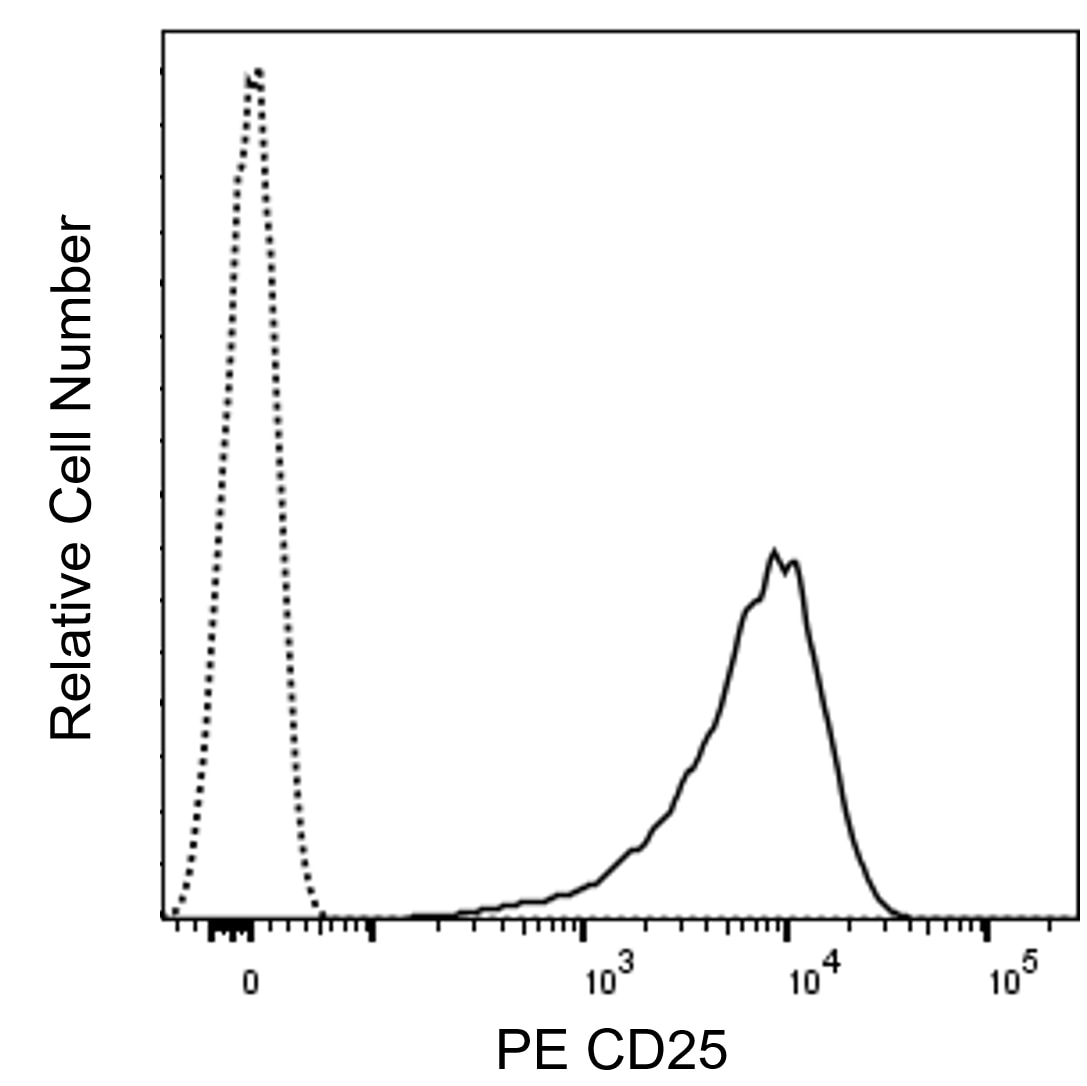
The BC96 monoclonal antibody specifically binds to human CD25, the low-affinity alpha subunit of the Interleukin-2 Receptor (IL-2Rα), which is also known as TAC antigen. IL2RA encodes CD25 which is a 55 kDa type I transmembrane glycoprotein comprised of an extracellular region with two Complement Control Protein domains (CCP) followed by a transmembrane region and a short cytoplasmic tail. CD25 is constitutively expressed at high levels on natural T regulatory cells and variably expressed on conventional T cells and B cells and their precursors, NK cells, monocytes, and macrophages. CD25 expression can be highly upregulated upon antigenic or mitogenic stimulation of T cells or B cells. A soluble form of CD25 is found in biological fluids due to proteolytic cleavage of the extracellular region of transmembrane CD25. CD25 noncovalently associates with CD122 (IL-2Rβ chain) and CD132 (IL-2Rγ, also known as the common γ chain or γc) to form the high-affinity signal-transducing IL-2R complex (IL-2Rαβγ).  This heterotrimeric receptor mediates biological activities of IL-2 which can act as a cellular activation, growth, and differentiation factor and regulator of cell viability. Analysis of CD25 expression can be used to characterize the nature of normal leucocytes in their resting states or activated during inflammatory or immune responses as well as those present in certain disease states.

567214 Rev. 2
Format Details
Down Arrow Up Arrow
PE
R-Phycoerythrin (PE), is part of the BD family of Phycobiliprotein dyes. This fluorochrome is a multimeric fluorescent phycobiliprotein with excitation maximum (Ex Max) of 496 nm and 566 nm and an emission maximum (Em Max) at 576 nm. PE is designed to be excited by the Blue (488 nm), Green (532 nm) and Yellow-Green (561 nm) lasers and detected using an optical filter centered near 575 nm (e.g., a 575/26-nm bandpass filter). As PE is excited by multiple lasers, this can result in cross-laser excitation and fluorescence spillover on instruments with various combinations of Blue, Green, and Yellow-Green lasers. Please ensure that your instrument’s configurations (lasers and optical filters) are appropriate for this dye.
altImg
PE
Yellow-Green 488 nm, 532 nm, 561 nm
496 nm, 566 nm
576 nm
567214 Rev.2
Citations & References
Down Arrow Up Arrow

Development References (4)

  1. Chapel A, Bensussan A, Vilmer E, Dormont D. Differential human immunodeficiency virus expression in CD4+ cloned lymphocytes: from viral latency to replication.. J Virol. 1992; 66(6):3966-70. (Immunogen: Flow cytometry). View Reference
  2. Knapp W. W. Knapp .. et al., ed. Leucocyte typing IV : white cell differentiation antigens. Oxford New York: Oxford University Press; 1989:1-1182.
  3. Poszepczynska E, Bagot M, Echchakir H, et al. Functional characterization of an IL-7-dependent CD4(+)CD8alphaalpha(+) Th3-type malignant cell line derived from a patient with a cutaneous T-cell lymphoma.. 2000; 96(3):1056-63. (Clone-specific). View Reference
  4. Schlossman SF. Stuart F. Schlossman .. et al., ed. Leucocyte typing V : white cell differentiation antigens : proceedings of the fifth international workshop and conference held in Boston, USA, 3-7 November, 1993. Oxford: Oxford University Press; 1995.
View All (4) View Less
567214 Rev. 2

Please refer to Support Documents for Quality Certificates


Global - Refer to manufacturer's instructions for use and related User Manuals and Technical data sheets before using this products as described


Comparisons, where applicable, are made against older BD Technology, manual methods or are general performance claims.  Comparisons are not made against non-BD technologies, unless otherwise noted.

For Research Use Only. Not for use in diagnostic or therapeutic procedures.