

Preparation And Storage
Recommended Assay Procedures
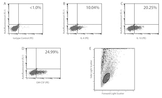
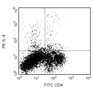
1. Blocking Control for Intracellular Staining: The purified BVD4-1D11 antibody (Cat. No. 554386) can be used as a blocking control to demonstrate specificity of IL-4 staining by conjugated-BVD4-1D11 and antibody. To perform this control, the fixed/permeabilized cells (~ 1 million) can be incubated with 1-10 µg of unlabeled BVD4-1D11 antibody (Cat. No. 554386) for 20 minutes at 4°C, prior to staining with conjugated-BVD4-1D11 antibody (e.g., 0.1 -0.5 µg mAb/1 million cells). The intracellular cytokine staining technique and use of blocking controls are described in detail by C. Prussin and D. Metcalfe. For specific methodology, please visit our web site, www.bdbiosciences.com, and go to the protocols section or the chapter on intracellular staining in the Immune Function Handbook.
2. ELISA Capture: The purified BVD4-1D11 antibody (Cat. No. 554387) is useful as a capture antibody for a sandwich ELISA for measuring mouse IL-4 protein levels in tissue culture supernatants. This antibody can be paired with the biotinylated BVD6-24G2 antibody (Cat. No. 554390) and recombinant mouse IL-4 (Cat. No. 550067) as the standard. For detecting IL-4 in serum or plasma, the BD OptEIA™ Mouse IL-4 ELISA Set (Cat. No. 555232) is recommended.
Note: Purified BVD4-1D11 antibody has been found to yield higher background in ELISA than the alternative mouse IL-4 ELISA capture antibody, clone 11B11 (Cat. No. 554434). To overcome this high background, the following procedures specific for clone BVD4-1D11 are recommended: 1) Titrate BVD4-1D11 capture antibody (1 -4 µg/ml) versus biotinylated BVD6-24G2 detection antibody (0.1 - 1.0 µg/ml; i.e. lower than usual detection antibody concentration). Separate blanks must be used for each concentration of biotinylated detection antibody used. 2) Use twice the number of recommended washes at all steps of ELISA protocol.
3. Neutralization: The NA/LE™ BVD4-1D11 antibody (Cat. No. 554385) is useful for neutralization of mouse IL-4 bioactivity.
4. WB: The BVD4-1D11 antibody has been reported to be useful for Western blotting. Please note that this application is not routinely tested at BD Biosciences Pharmingen.
Product Notices
- Since applications vary, each investigator should titrate the reagent to obtain optimal results.
- Please refer to www.bdbiosciences.com/us/s/resources for technical protocols.
- Caution: Sodium azide yields highly toxic hydrazoic acid under acidic conditions. Dilute azide compounds in running water before discarding to avoid accumulation of potentially explosive deposits in plumbing.
Companion Products
The BVD4-1D11 antibody reacts with mouse interleukin-4 (IL-4). The immunogen used to generate the BVD4-1D11 hybridoma was recombinant mouse IL-4.
This antibody is routinely tested by intracellular staining and sandwich ELISA. Other applications were tested at BD Biosciences Pharmingen during antibody development only or reported in the literature.
Development References (5)
-
Abrams JS, Roncarolo MG, Yssel H, Andersson U, Gleich GJ, Silver JE. Strategies of anti-cytokine monoclonal antibody development: immunoassay of IL-10 and IL-5 in clinical samples. Immunol Rev. 1992; 127:5-24. (Clone-specific: ELISA, Neutralization). View Reference
-
Finkelman FD, Madden KB, Morris SC, et al. Anti-cytokine antibodies as carrier proteins. Prolongation of in vivo effects of exogenous cytokines by injection of cytokine-anti-cytokine antibody complexes. J Immunol. 1993; 151(3):1235-1244. (Clone-specific: ELISA, Neutralization). View Reference
-
Litton MJ, Sander B, Murphy E, O'Garra A, Abrams JS. Early expression of cytokines in lymph nodes after treatment in vivo with Staphylococcus enterotoxin B. J Immunol Methods. 1994; 175(1):47-58. (Clone-specific: ELISA). View Reference
-
Prussin C, Metcalfe DD. Detection of intracytoplasmic cytokine using flow cytometry and directly conjugated anti-cytokine antibodies. J Immunol Methods. 1995; 188(1):117-128. (Clone-specific: IC/FCM Block). View Reference
-
Sander B, Hoiden I, Andersson U, Moller E, Abrams JS. Similar frequencies and kinetics of cytokine producing cells in murine peripheral blood and spleen. Cytokine detection by immunoassay and intracellular immunostaining. J Immunol Methods. 1993; 166(2):201-214. (Clone-specific: ELISA, Neutralization). View Reference
Please refer to Support Documents for Quality Certificates
Global - Refer to manufacturer's instructions for use and related User Manuals and Technical data sheets before using this products as described
Comparisons, where applicable, are made against older BD Technology, manual methods or are general performance claims. Comparisons are not made against non-BD technologies, unless otherwise noted.
For Research Use Only. Not for use in diagnostic or therapeutic procedures.