![Biotin Mouse Anti-Mouse H-2K[b]](/content/dam/bdb/products/global/reagents/flow-cytometry-reagents/research-reagents/single-color-antibodies-ruo/550xxx/5505xx/550550_base/75192E_550550_image1.png)
Preparation And Storage
Recommended Assay Procedures
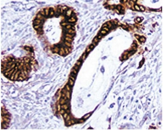
The AF6-88.5 clone reactive against mouse H-2Kb is recommended to test for immunohistorical staining of acetone-fixed frozen sections. Tissues tested were mouse (C57B1/6) spleen and thymus. The antibody stains the H-2Kb molecules found on the surface of all nucleated cells in strains of mice expressing this haplotype. For optimal immunohistochemical staining, the biotinylated antibody should be titrated (1:10 to 1:50 dilution) and developed with Streptavidin-HRP (Cat. No. 550946) together with the DAB detection system (Cat. No. 550880). The clone AF6-88.5 is not recommended for formalin-fixed paraffin embedded sections.
Product Notices
- Since applications vary, each investigator should titrate the reagent to obtain optimal results.
- Source of all serum proteins is from USDA inspected abattoirs located in the United States.
- Caution: Sodium azide yields highly toxic hydrazoic acid under acidic conditions. Dilute azide compounds in running water before discarding to avoid accumulation of potentially explosive deposits in plumbing.
- An isotype control should be used at the same concentration as the antibody of interest.
- This antibody has been developed for the immunohistochemistry application. However, a routine immunohistochemistry test is not performed on every lot. Researchers are encouraged to titrate the reagent for optimal performance.
- For fluorochrome spectra and suitable instrument settings, please refer to our Multicolor Flow Cytometry web page at www.bdbiosciences.com/colors.
- Please refer to www.bdbiosciences.com/us/s/resources for technical protocols.
Companion Products


The AF6-88.5 antibody recognizes the H-2Kb MHC class I alloantigen. Reactivity with other haplotypes (e.g., d, f, j, k, p, q, r, s, u, v) has not been observed.
Development References (2)
-
Loken MR, Stall AM. Flow cytometry as an analytical and preparative tool in immunology. J Immunol Methods. 1982; 50(3):R85-R112. (Immunogen). View Reference
-
Wall KA, Lorber MI, Loken MR, McClatchey S, Fitch FW. Inhibition of proliferation of MIs- and Ia-reactive cloned T cells by a monoclonal antibody against a determinant shared by I-A and I-E.. J Immunol. 1983; 131(3):1056-64. (Biology). View Reference
Please refer to Support Documents for Quality Certificates
Global - Refer to manufacturer's instructions for use and related User Manuals and Technical data sheets before using this products as described
Comparisons, where applicable, are made against older BD Technology, manual methods or are general performance claims. Comparisons are not made against non-BD technologies, unless otherwise noted.
For Research Use Only. Not for use in diagnostic or therapeutic procedures.