
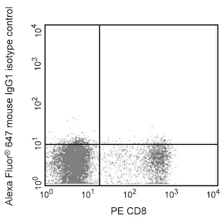
Flow cytometric analysis of IFN-γ expression by stimulated CD4+ rat splenocytes. Purified splenic CD4+ cells from Lewis rats were stimulated with plate-bound Purified NA/LE Mouse Anti-Rat CD3 (10 μg/ml; Cat. No. 554829) and soluble Purified NA/LE Mouse Anti-Rat CD28 (2 μg/ml; Cat. No. 554993) for 2 days in complete medium with Recombinant Rat IL-2 (10 ng/ml; Cat. No. 555106) and Recombinant Rat IL-4 (40 ng/ml; Cat. No 555107). Cells were harvested and cultured for 3 days in the presence of IL-2 (10 ng/ml) and IL-4 (20 ng/ml). This was followed by a 6 hour stimulation with Phorbol 12-Myristate 13-Acetate (PMA; 5 ng/ml; Sigma, Cat. #P-8139) and Ionomycin (500 ng/ml; Sigma, Cat. #I-0634) in the presence of GolgiPlug™ Protein Transport Inhibitor (Containing Brefeldin A) (2 μM; Cat. No. 555029). The cells were then fixed with BD Cytofix™ Fixation Buffer (Cat. No. 554655), and permeabilized with BD Perm/Wash™ Buffer (Cat. No. 554723), and subsequently stained with either Alexa Fluor® 647 IgG1, κ Isotype Control (Cat. No. 557732, Left Panel) or Alexa Fluor® 647 Mouse Anti-Rat IFN-γ antibody (Middle and Right Panel, Cat. No. 562213) by using BD Biosciences Intracellular Cytokine Staining Protocol. To demonstrate the specificity of staining, the binding of the Alexa Fluor® 647 Mouse Anti-Rat IFN-γ antibody was blocked by preincubation of cells with the Purified Mouse Anti-Rat IFN-γ antibody (Clone DB-1; Right Panel) prior to staining. Flow cytometric fluorescence dot blots showing the expression of IFN-γ (or Ig Isotype background staining) versus cellular autofluorescence (measured in the FL2 channel) were derived from gated events with the forward and side light-scatter characteristics of intact splenocytes. Flow cytometry was performed using a BD™ LSR II Flow Cytometer System.

Flow cytometric analysis of IFN-γ expression by stimulated CD4+ rat splenocytes. Purified splenic CD4+ cells from Lewis rats were stimulated with plate-bound Purified NA/LE Mouse Anti-Rat CD3 (10 μg/ml; Cat. No. 554829) and soluble Purified NA/LE Mouse Anti-Rat CD28 (2 μg/ml; Cat. No. 554993) for 2 days in complete medium with Recombinant Rat IL-2 (10 ng/ml; Cat. No. 555106) and Recombinant Rat IL-4 (40 ng/ml; Cat. No 555107). Cells were harvested and cultured for 3 days in the presence of IL-2 (10 ng/ml) and IL-4 (20 ng/ml). This was followed by a 6 hour stimulation with Phorbol 12-Myristate 13-Acetate (PMA; 5 ng/ml; Sigma, Cat. #P-8139) and Ionomycin (500 ng/ml; Sigma, Cat. #I-0634) in the presence of GolgiPlug™ Protein Transport Inhibitor (Containing Brefeldin A) (2 μM; Cat. No. 555029). The cells were then fixed with BD Cytofix™ Fixation Buffer (Cat. No. 554655), and permeabilized with BD Perm/Wash™ Buffer (Cat. No. 554723), and subsequently stained with either Alexa Fluor® 647 IgG1, κ Isotype Control (Cat. No. 557732, Left Panel) or Alexa Fluor® 647 Mouse Anti-Rat IFN-γ antibody (Middle and Right Panel, Cat. No. 562213) by using BD Biosciences Intracellular Cytokine Staining Protocol. To demonstrate the specificity of staining, the binding of the Alexa Fluor® 647 Mouse Anti-Rat IFN-γ antibody was blocked by preincubation of cells with the Purified Mouse Anti-Rat IFN-γ antibody (Clone DB-1; Right Panel) prior to staining. Flow cytometric fluorescence dot blots showing the expression of IFN-γ (or Ig Isotype background staining) versus cellular autofluorescence (measured in the FL2 channel) were derived from gated events with the forward and side light-scatter characteristics of intact splenocytes. Flow cytometry was performed using a BD™ LSR II Flow Cytometer System.
Product Details


BD Pharmingen™
Ifng; IFN-g; IFNγ; IFN-γ; IFNG2; IFN-gamma; Interferon gamma
Rat (QC Testing)
Mouse IgG1, κ
Recombinant Rat IFN-γ
Intracellular staining (flow cytometry) (Routinely Tested)
5 µl
25712
AB_11153686
Aqueous buffered solution containing BSA and ≤0.09% sodium azide.
RUO
Preparation And Storage
Store undiluted at 4°C and protected from prolonged exposure to light. Do not freeze. The monoclonal antibody was purified from tissue culture supernatant or ascites by affinity chromatography. The antibody was conjugated to Alexa Fluor® 647 under optimum conditions, and unreacted Alexa Fluor® 647 was removed.
Product Notices
- Please refer to www.bdbiosciences.com/us/s/resources for technical protocols.
- Since applications vary, each investigator should titrate the reagent to obtain optimal results.
- The Alexa Fluor®, Pacific Blue™, and Cascade Blue® dye antibody conjugates in this product are sold under license from Molecular Probes, Inc. for research use only, excluding use in combination with microarrays, or as analyte specific reagents. The Alexa Fluor® dyes (except for Alexa Fluor® 430), Pacific Blue™ dye, and Cascade Blue® dye are covered by pending and issued patents.
- Alexa Fluor® is a registered trademark of Molecular Probes, Inc., Eugene, OR.
- Alexa Fluor® 647 fluorochrome emission is collected at the same instrument settings as for allophycocyanin (APC).
- Caution: Sodium azide yields highly toxic hydrazoic acid under acidic conditions. Dilute azide compounds in running water before discarding to avoid accumulation of potentially explosive deposits in plumbing.
- For fluorochrome spectra and suitable instrument settings, please refer to our Multicolor Flow Cytometry web page at www.bdbiosciences.com/colors.
- Source of all serum proteins is from USDA inspected abattoirs located in the United States.
- An isotype control should be used at the same concentration as the antibody of interest.
Companion Products
Stain Buffer (FBS) RUO
Size
500 mL
Cat No.
554656

Fixation Buffer RUO
Size
100 mL
Cat No.
554655

Perm/Wash Buffer RUO
Size
100 mL
Cat No.
554723

Alexa Fluor® 647 Mouse IgG1 κ Isotype Control RUO
Size
100 Tests
Cat No.
557732
Protein Transport Inhibitor (Containing Brefeldin A) RUO
Size
1 mL
Cat No.
555029
Purified NA/LE Mouse Anti-Rat CD3 RUO
Size
0.5 mg
Cat No.
554829

562213 Rev. 1
Antibody Details
DB-1
The DB-1 monoclonal antibody specifically binds to rat interferon-γ (IFN-γ). The immunogen used to generate the DB-1 hybridoma was recombinant rat IFN-γ expressed in COS cells. This is a neutralizing antibody.
562213 Rev. 1
Format Details
Alexa Fluor™ 647
Alexa Fluor™ 647 Dye is part of the BD red family of dyes. This is a small organic fluorochrome with an excitation maximum (Ex Max) at 653-nm and an emission maximum (Em Max) at 669-nm. Alexa Fluor 647 is designed to be excited by the Red laser (627-640 nm) and detected using an optical filter centered near 520-nm (e.g., a 660/20 nm bandpass filter). Please ensure that your instrument’s configurations (lasers and optical filters) are appropriate for this dye.
Alexa Fluor™ 647
Red 627-640 nm
653 nm
669 nm
562213 Rev.1
Citations & References
Development References (7)
-
Bakhiet M, Olsson T, Mhlanga J. Human and rodent interferon-gamma as a growth factor for Trypanosoma brucei. Eur J Immunol. 1996; 26(6):1359-1364. (Biology). View Reference
-
Prussin C, Metcalfe DD. Detection of intracytoplasmic cytokine using flow cytometry and directly conjugated anti-cytokine antibodies. J Immunol Methods. 1995; 188(1):117-128. (Methodology: Flow cytometry). View Reference
-
Schmidt B, Stoll G, van der Meide P, Jung S, Hartung HP. Transient cellular expression of gamma-interferon in myelin-induced and T-cell line-mediated experimental autoimmune neuritis. 1992; 115(Pt 6):1633-1646. (Biology). View Reference
-
van der Meide PH, Borman AH, Beljaars HG, Dubbeld MA, Botman CA, Schellekens H. Isolation and characterization of monoclonal antibodies directed to rat interferon-gamma. Leuk Res. 1989; 8(4):439-449. (Biology). View Reference
-
van der Meide PH, Borman TH, de Labie MC, et al. A sensitive two-site enzyme immunoassay for the detection of rat interferon-gamma in biological fluids. J Interferon Res. 1990; 10(2):183-189. (Biology). View Reference
-
van der Meide PH, Dubbeld M, Vijverberg K, Kos T, Schellekens H. The purification and characterization of rat gamma interferon by use of two monoclonal antibodies.. J Gen Virol. 1986; 67(Pt 6):1059-1071. (Biology). View Reference
-
van der Meide PH, Groenestein RJ, de Labie MC, Aten J, Weening JJ. Susceptibility to mercuric chloride-induced glomerulonephritis is age-dependent: study of the role of IFN-gamma. Cell Immunol. 1995; 162(1):131-137. (Biology). View Reference
562213 Rev. 1
Please refer to Support Documents for Quality Certificates
Global - Refer to manufacturer's instructions for use and related User Manuals and Technical data sheets before using this products as described
Comparisons, where applicable, are made against older BD Technology, manual methods or are general performance claims. Comparisons are not made against non-BD technologies, unless otherwise noted.
For Research Use Only. Not for use in diagnostic or therapeutic procedures.