







Preparation And Storage


Recommended Assay Procedures
Bioimaging
1. Seed the cells in appropriate culture medium at ~10,000 cells per well in a BD Falcon™ 96-well Imaging Plate (Cat. No. 353219) and culture overnight.
2. Remove the culture medium from the wells, and fix the cells by adding 100 μl of BD Cytofix™ Fixation Buffer (Cat. No. 554655) to each well. Incubate for 10 minutes at room temperature (RT).
3. Remove the fixative from the wells, and permeabilize the cells using either BD Perm Buffer III, 90% methanol, or Triton™ X-100:
a. Add 100 μl of -20°C 90% methanol or Perm Buffer III (Cat. No. 558050) to each well and incubate for 5 minutes at RT.
OR
b. Add 100 μl of 0.1% Triton™ X-100 to each well and incubate for 5 minutes at RT.
4. Remove the permeabilization buffer, and wash the wells twice with 100 μl of 1× PBS.
5. Remove the PBS, and block the cells by adding 100 μl of BD Pharmingen™ Stain Buffer (FBS) (Cat. No. 554656) to each well. Incubate for 30 minutes at RT.
6. Remove the blocking buffer and add 50 μl of the optimally titrated primary antibody (diluted in Stain Buffer) to each well, and incubate for 1 hour at RT.
7. Remove the primary antibody, and wash the wells three times with 100 μl of 1× PBS.
8. Remove the PBS, and add the second step reagent at its optimally titrated concentration in 50 μl to each well, and incubate in the dark for 1 hour at RT.
9. Remove the second step reagent, and wash the wells three times with 100 μl of 1× PBS.
10. Remove the PBS, and counter-stain the nuclei by adding 200 μl per well of 2 μg/ml Hoechst 33342 (e.g., Sigma-Aldrich Cat. No. B2261) in 1× PBS to each well at least 15 minutes before imaging.
11. View and analyze the cells on an appropriate imaging instrument.
Bioimaging: For more detailed information please refer to http://www.bdbiosciences.com/support/resources/protocols/ceritifed_reagents.jsp
Western blot: For more detailed information please refer to http://www.bdbiosciences.com/pharmingen/protocols/Western_Blotting.shtml
Product Notices
- Since applications vary, each investigator should titrate the reagent to obtain optimal results.
- Please refer to www.bdbiosciences.com/us/s/resources for technical protocols.
- This antibody has been developed and certified for the bioimaging application. However, a routine bioimaging test is not performed on every lot. Researchers are encouraged to titrate the reagent for optimal performance.
- Caution: Sodium azide yields highly toxic hydrazoic acid under acidic conditions. Dilute azide compounds in running water before discarding to avoid accumulation of potentially explosive deposits in plumbing.
- Source of all serum proteins is from USDA inspected abattoirs located in the United States.
- Triton is a trademark of the Dow Chemical Company.
Companion Products




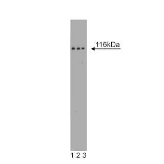
Focal Adhesion Kinase (FAK) is a cytoplasmic tyrosine kinase that colocalizes with integrins in focal adhesions. This cellular localization is directed by a 125 amino acid sequence at the C-terminus called the "Focal Adhesion Targeting" sequence (FAT). The binding of extracellular matrix ligands to integrins triggers autophosphorylation at Tyr-397, and activation of FAK through phosphorylation of Tyr residues (Tyr-576 and Tyr577) in the kinase domain activation loop. For example, cell adhesion to a fibronectin substratum involves concurrent activation of Src and phosphorylation of the FAK activation loop. In addition, phosphorylation of other Tyr residues (Tyr-925, and Tyr-861) creates binding sites for SH2 domains of intracellular signaling molecules such as Src, PI3 kinase, and Grb2. FAK's ability to bind numerous structural and signaling proteins via a variety of interactions is important for FAK activation level, and for FAK interaction with a variety of substrates localized to sites of cell adhesion. Thus, FAK activity is regulated by a complex set of phosphorylation sites, and this phospho-regulation could be important for cell motility, cell growth, cytoskeletal organization, and adhesion-dependent cell survival.
Development References (3)
-
Calalb MB, Zhang X, Polte TR, Hanks SK. Focal adhesion kinase tyrosine-861 is a major site of phosphorylation by Src. Biochem Biophys Res Commun. 1996; 228(3):662-668. (Biology). View Reference
-
McLean GW, Fincham VJ, Frame MC. v-Src induces tyrosine phosphorylation of focal adhesion kinase independently of tyrosine 397 and formation of a complex with Src. J Biol Chem. 2000; 275(30):23333-23339. (Biology). View Reference
-
Ruest PJ, Roy S, Shi E, Mernaugh RL, Hanks SK. Phosphospecific antibodies reveal focal adhesion kinase activation loop phosphorylation in nascent and mature focal adhesions and requirement for the autophosphorylation site. Cell Growth Differ. 2000; 11(1):41-48. (Biology). View Reference
Please refer to Support Documents for Quality Certificates
Global - Refer to manufacturer's instructions for use and related User Manuals and Technical data sheets before using this products as described
Comparisons, where applicable, are made against older BD Technology, manual methods or are general performance claims. Comparisons are not made against non-BD technologies, unless otherwise noted.
For Research Use Only. Not for use in diagnostic or therapeutic procedures.
