BD Pharmingen™ Alexa Fluor® 647 Mouse Anti-Human MUC1 (CD227)
Clone HMFG2 (also known as HMFG-2, 3.14.A3 or F314AE11)
(RUO)

Preparation And Storage
Product Notices
- Since applications vary, each investigator should titrate the reagent to obtain optimal results.
- An isotype control should be used at the same concentration as the antibody of interest.
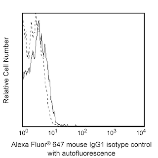
- Alexa Fluor® 647 fluorochrome emission is collected at the same instrument settings as for allophycocyanin (APC).
- Alexa Fluor® is a registered trademark of Molecular Probes, Inc., Eugene, OR.
- The Alexa Fluor®, Pacific Blue™, and Cascade Blue® dye antibody conjugates in this product are sold under license from Molecular Probes, Inc. for research use only, excluding use in combination with microarrays, or as analyte specific reagents. The Alexa Fluor® dyes (except for Alexa Fluor® 430), Pacific Blue™ dye, and Cascade Blue® dye are covered by pending and issued patents.
- Caution: Sodium azide yields highly toxic hydrazoic acid under acidic conditions. Dilute azide compounds in running water before discarding to avoid accumulation of potentially explosive deposits in plumbing.
- For fluorochrome spectra and suitable instrument settings, please refer to our Multicolor Flow Cytometry web page at www.bdbiosciences.com/colors.
- Please refer to www.bdbiosciences.com/us/s/resources for technical protocols.
Companion Products


The HMFG2 (Milk Fat Globule 2) monoclonal antibody, formerly known as clone 3.14.A3, specifically binds to Mucin-1 (MUC1) which is also known as CD227, Episialin, Breast carcinoma-associated antigen DF3, Cancer antigen 15-3, H23 antigen, Peanut reactive urinary protein (PUM), Polymorphic epithelial mucin (PEM), or Epithelial membrane antigen (EMA). MUC1 (CD227) is a heterodimeric cell surface glycoprotein that belongs to the epithelial mucin family. As a result of proteolytic cleavage, MUC1 (CD227) is comprised of a large, extracellular N-terminal alpha subunit that contains a domain of 20 amino-acid tandem repeats and functions in cellular adhesion. The smaller transmembrane C-terminal beta subunit contains a cytoplasmic domain that is involved in cell signaling. MUC1 (CD227) is variably expressed on the surfaces of normal and malignant glandular and ductal epithelial cells, myeloma cells, and some hematopoietic cell lineages including subsets of T cells, B cells, monocytes and dendritic cells. Soluble forms of MUC1 (CD227) may arise by shedding from the cell surface or by secretion of forms derived from alternative RNA splicing. MUC1 (CD227) plays roles in the provision of protective barrier function, the regulation of cellular adhesion and migration, and the transduction of multiple signal pathways. The HMFG2 antibody recognizes epitopes within the tandem repeat domain of MUC1 (CD227).
Development References (7)
-
Arklie J, Taylor-Papadimitrious J, Bodmer W, Egan M, Millis R. Differentiation antigens expressed by epithelial cells in the lactating breast are also detectable in breast cancers.. Int J Cancer. 1981; 28(1):23-9. (Clone-specific: Immunohistochemistry). View Reference
-
Burchell J, Durbin H, Taylor-Papadimitriou J. Complexity of expression of antigenic determinants, recognized by monoclonal antibodies HMFG-1 and HMFG-2, in normal and malignant human mammary epithelial cells.. J Immunol. 1983; 131(1):508-13. (Clone-specific: Radioimmunoassay, Western blot). View Reference
-
Burchell J, Taylor-Papadimitriou J. Effect of modification of carbohydrate side chains on the reactivity of antibodies with core-protein epitopes of the MUC1 gene product.. Epithelial Cell Biol. 1993; 2(4):155-62. (Clone-specific: Flow cytometry, Immunohistochemistry). View Reference
-
Correa I, Plunkett T, Vlad A, et al. Form and pattern of MUC1 expression on T cells activated in vivo or in vitro suggests a function in T-cell migration.. Immunology. 2003; 108(1):32-41. (Clone-specific: Flow cytometry, Fluorescence microscopy, Immunofluorescence). View Reference
-
McGuckin MA, MacDonald KP, Tran M, Wykes M, Hart DNJ. MUC1 Epithelial mucin: expression by normal hematopoietic cells. In: Mason D. David Mason .. et al., ed. Leucocyte typing VII : white cell differentiation antigens : proceedings of the Seventh International Workshop and Conference held in Harrogate, United Kingdom. Oxford: Oxford University Press; 2002:496-499.
-
McGuckin MA. CD227 (MUC1) Summary and Workshop Report. In: Mason D. David Mason .. et al., ed. Leucocyte typing VII : white cell differentiation antigens : proceedings of the Seventh International Workshop and Conference held in Harrogate, United Kingdom. Oxford: Oxford University Press; 2002:494-496.
-
Taylor-Papadimitriou J, Peterson JA, Arklie J, Burchell J, Ceriani RL, Bodmer WF. Monoclonal antibodies to epithelium-specific components of the human milk fat globule membrane: production and reaction with cells in culture.. Int J Cancer. 1981; 28(1):17-21. (Immunogen: Immunoprecipitation, Radioimmunoassay). View Reference
Please refer to Support Documents for Quality Certificates
Global - Refer to manufacturer's instructions for use and related User Manuals and Technical data sheets before using this products as described
Comparisons, where applicable, are made against older BD Technology, manual methods or are general performance claims. Comparisons are not made against non-BD technologies, unless otherwise noted.
For Research Use Only. Not for use in diagnostic or therapeutic procedures.