Skip to main content Skip to navigation
Alexa Fluor® 488 Mouse anti-Human Bcl-2 (pS70)
Flow cytometric (Panels 1a and 1b) and Western blot (Panel 1c) analyses of Bcl-2 (pS70) expressed by Jurkat cells. Jurkat cells were either not treated (Panel 1a, dashed line histogram; Panel 1c, C) or treated (Panel 1a, solid line histogram; Panel 1b; Panel 1c, T) with 100 nM Taxol (Sigma, Cat. No. T7191; 24 hr, 37°C). For Panel 1a, cells were fixed in BD Phosflow™ Cytofix Buffer (Cat. No. 554655; 10 min, 37˚C) and permeabilized in BD Phosflow™ Perm Buffer III (Cat. No. 558050; 30 min, on ice) prior to staining with Alexa Fluor® 488 Mouse Anti-Bcl-2 (pS70) antibody (Cat. No. 562678). For optimal co-staining of total cellular DNA (Panel 1b), cells were fixed and permeabilized in 70% ethanol (30 min, on ice) prior to staining with 7-AAD (Cat. No. 559925) and Alexa Fluor® 488 Mouse Anti-Bcl-2 (pS70). Fluorescence histograms (Panel 1a) and a two-color dot plot showing DNA (7-AAD) versus Bcl-2 (pS70) levels (Panel 1b) were generated from gated events with the light scatter characteristics of intact cells using a BD™ LSR II Flow Cytometer System. For Western blot analysis (Panel 1c), cell lysates (15 µg total cell protein/lane) were blotted using Purified Mouse Anti-Bcl-2 (pS70) (Cat. No. 562529; 0.125, 0.063, and 0.032 µg/ml for Lanes 1, 2, 3, respectively). Bcl-2 (pS70) was identified as a ~28 kDa band. Flow cytometric (Panels 2a and 2b) and Western blot (Panel 2c) analyses of Bcl-2 (pS70) expressed by human peripheral blood mononuclear cells. PHA-stimulated (20 μg/ml for 3 days; Sigma Cat. No. L1668) PBMC were either not treated (Panel 2a, dashed line histogram; Panel 2c, C) or treated (Panel 2a, solid line histogram; Panel 2b; Panel 2c, T) with Taxol (100 nM, 24 hr, 37°C). Flow cytometric analyses of Bcl-2 (pS70) expression without (Panel 2a) or with (Panel 2b) co-staining for DNA content were performed using a BD FACSCanto™ II Flow Cytometer System. Lysates from 1X10^6 PBMC were blotted using Purified Mouse Anti-Bcl-2 (pS70) antibody (2.0 µg/ml) as described.
Product Details
Down Arrow Up Arrow


BD Phosflow™
BCL2; Apoptosis regulator Bcl-2; B-cell CLL/lymphoma 2; PPP1R50
Human (QC Testing), Mouse (Tested in Development)
Mouse IgG1
Phosphorylated Peptide
Intracellular staining (flow cytometry) (Routinely Tested)
5 µl
AB_2737718
Aqueous buffered solution containing BSA and ≤0.09% sodium azide.
RUO


Preparation And Storage

Store undiluted at 4°C and protected from prolonged exposure to light. Do not freeze. The monoclonal antibody was purified from tissue culture supernatant or ascites by affinity chromatography. The antibody was conjugated to Alexa Fluor® 488 under optimum conditions, and unreacted Alexa Fluor® 488 was removed.

Product Notices

  1. This reagent has been pre-diluted for use at the recommended Volume per Test. We typically use 1 × 10^6 cells in a 100-µl experimental sample (a test).
  2. Source of all serum proteins is from USDA inspected abattoirs located in the United States.
  3. The Alexa Fluor®, Pacific Blue™, and Cascade Blue® dye antibody conjugates in this product are sold under license from Molecular Probes, Inc. for research use only, excluding use in combination with microarrays, or as analyte specific reagents. The Alexa Fluor® dyes (except for Alexa Fluor® 430), Pacific Blue™ dye, and Cascade Blue® dye are covered by pending and issued patents.
  4. Alexa Fluor® 488 fluorochrome emission is collected at the same instrument settings as for fluorescein isothiocyanate (FITC).
  5. Alexa Fluor® is a registered trademark of Molecular Probes, Inc., Eugene, OR.
  6. Caution: Sodium azide yields highly toxic hydrazoic acid under acidic conditions. Dilute azide compounds in running water before discarding to avoid accumulation of potentially explosive deposits in plumbing.
  7. For fluorochrome spectra and suitable instrument settings, please refer to our Multicolor Flow Cytometry web page at www.bdbiosciences.com/colors.
  8. All other brands are trademarks of their respective owners.
  9. Species cross-reactivity detected in product development may not have been confirmed on every format and/or application.
  10. Please refer to www.bdbiosciences.com/us/s/resources for technical protocols.
562678 Rev. 1
Antibody Details
Down Arrow Up Arrow
N46-467

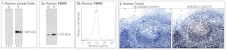
The N46-467 monoclonal antibody specifically binds to Bcl-2 (pS70), ie, the Bcl-2 protein phosphorylated at the Ser70 site. Bcl-2 is a ~ 26 kDa intracellular, integral membrane protein found primarily in the nuclear envelope, endoplasmic reticulum and outer mitochondrial membrane. Bcl-2 is encoded by the BCL2 (B-cell CLL/lymphoma 2) gene and is also known as Apoptosis regulator Bcl-2. Members of the Bcl-2 family play a major role in regulating the response of cells to apoptotic signals. Bcl-2 is one of the anti-apoptotic members of the Bcl-2 family. Bcl-2 knockout mice showed pronounced lymphoid apoptosis and other apoptosis related lesions later in life.  Bcl-2 is a proto-oncogene because it blocks apoptosis and provides a selective survival advantage in many cell types and thus contributes to tumorigenesis. It has been implicated in several types of cancers, such as breast, prostate, and melanoma . Bcl-2 contains multiple phosphorylation sites including Thr56, Ser70, Thr74 and Ser87. Phosphorylation of Bcl-2 Ser70 has been shown to be a mitotic marker. Phosphorylation at this site regulates Bcl-2's anti-apoptotic activity and has recently been implicated in promoting autophagy. Several studies have shown that Bcl-2 phosphorylation is caused by c-Jun N-terminal kinase (JNK).

562678 Rev. 1
Format Details
Down Arrow Up Arrow
Alexa Fluor™ 488
Alexa Fluor™ 488 Dye is part of the BD blue family of dyes. This is a small organic fluorochrome with an excitation maximum (Ex Max) at 494-nm and an emission maximum (Em Max) at 517-nm. Alexa Fluor™ 488 is designed to be excited by the Blue laser (488 nm) and detected using an optical filter centered near 520-nm (e.g., a 530/30-nm bandpass filter). Please ensure that your instrument’s configurations (lasers and optical filters) are appropriate for this dye.
Alexa Fluor™ 488
Blue 488 nm
494 nm
517 nm
562678 Rev.1
Citations & References
Down Arrow Up Arrow

Development References (6)

  1. Deng X, Xiao L, Lang W, et al. Novel role for JNK as a stress-activated Bcl2 kinase. J Biol Chem. 2001; 276(26):23681-23688. (Biology). View Reference
  2. Geng F, Tang L, Li Y, et al. Allyl isothiocyanate arrests cancer cells in mitosis, and mitotic arrest in turn leads to apoptosis via Bcl-2 protein phosphorylation. J Biol Chem. 2011; 286(37):32259-32267. (Biology). View Reference
  3. Ling YH, Tornos C, Perez-Soler R. Phosphorylation of Bcl-2 is a marker of M phase events and not a determinant of apoptosis. J Biol Chem. 1998; 273(30):18984-18991. (Biology). View Reference
  4. Maundrell K, Antonsson B, Magnenat E, et al. Bcl-2 undergoes phosphorylation by c-Jun N-terminal kinase/stress-activated protein kinases in the presence of the constitutively active GTP-binding protein Rac1. J Biol Chem. 1997; 272(40):25238-42. (Biology). View Reference
  5. Pattingre S, Bauvy C, Carpentier S, Levade T, Levine B, Codogno P.. Role of JNK1-dependent Bcl-2 phosphorylation in ceramide-induced macroautophagy. J Biol Chem. 2009; 284(5):2719-2728. (Biology). View Reference
  6. Sarkar S, Korolchuk VI, Renna M, et al. Complex inhibitory effects of nitric oxide on autophagy. Mol Cell. 2011; 43(1):19-32. (Biology). View Reference
View All (6) View Less
562678 Rev. 1

Please refer to Support Documents for Quality Certificates


Global - Refer to manufacturer's instructions for use and related User Manuals and Technical data sheets before using this products as described


Comparisons, where applicable, are made against older BD Technology, manual methods or are general performance claims.  Comparisons are not made against non-BD technologies, unless otherwise noted.

For Research Use Only. Not for use in diagnostic or therapeutic procedures.