Skip to main content Skip to navigation
Alexa Fluor® 647 Rat Anti-Mouse Zbtb7b (ThPok)
Flow cytometric analysis of Zbtb7b (ThPOK) expression in C57BL/6 mouse thymocytes and lymph node cells.        Thymocytes (Panel 1) or Lymph Node Cells (Panels 2 and 3) were stained with BD Horizon™ BV421 Rat Anti-Mouse CD4 (Cat No. 562891) and PE Rat Anti-Mouse CD8a (Cat. No. 553033/553032/561095) antibodies. The cells were fixed and permeabilized with the BD Pharmingen™ Transcription Factor Buffer Set (Cat. No. 562574/562725) and stained with either Alexa Fluor® 647 Rat IgG2b, κ Isotype Control (Cat. No. 557691) or Alexa Fluor® 647 Rat Anti-Mouse Zbtb7b (ThPOK) (Cat. No. 565500) antibody. Two-color flow cytometric contour plots showing the correlated expression of Zbtb7b (ThPOK) [or Ig Isotype control staining] versus CD8a (Panel 1, Thymocytes) or CD4 (Panel 2, Lymph Node Cells) were derived from gated events with the forward and side light-scatter characteristics of intact cells. The fluorescence histograms (Panel 3, Lymph Node Cells) showing Zbtb7b (ThPOK) expression in CD4+ (solid line histogram) versus CD8+ cells (dashed line histogram) were derived from CD4+CD8- or CD4-CD8+ gated events, respectively, with the light-scatter characteristics of intact lymphocytes.        Alternatively, Lymph Node Cells (Panel 4) were similarly fixed, permeabilized, and stained with PE Mouse Anti-RUNX3 (Cat. No. 564814) and Alexa Fluor® 647 Rat Anti-Mouse Zbtb7b (ThPOK) antibodies. The two-color flow cytometric contour plot showing the correlated expression of Zbtb7b (ThPOK) versus RUNX3 was derived from gated events with the light-scatter characteristics of intact lymphocytes.        Flow cytometric analysis was performed using a BD LSRFortessa™ Flow Cytometer System.
Product Details
Down Arrow Up Arrow


BD Pharmingen™
Zbtb7b; ZBT7B; Th-POK; Thpok; c-Krox; Zfp-67; Zfp67
Mouse (QC Testing)
Rat WI, also known as Wistar (outbred) IgG2b, κ
Mouse Zbtb7b
Intracellular staining (flow cytometry) (Routinely Tested)
5 µl/test
AB_2739268
Aqueous buffered solution containing BSA and ≤0.09% sodium azide.
RUO


Preparation And Storage

Store undiluted at 4°C and protected from prolonged exposure to light. Do not freeze. The monoclonal antibody was purified from tissue culture supernatant or ascites by affinity chromatography. The antibody was conjugated to Alexa Fluor® 647 under optimum conditions, and unreacted Alexa Fluor® 647 was removed.

Product Notices

  1. This reagent has been pre-diluted for use at the recommended Volume per Test. We typically use 1 × 10^6 cells in a 100-µl experimental sample (a test).
  2. Please refer to www.bdbiosciences.com/us/s/resources for technical protocols.
  3. The Alexa Fluor®, Pacific Blue™, and Cascade Blue® dye antibody conjugates in this product are sold under license from Molecular Probes, Inc. for research use only, excluding use in combination with microarrays, or as analyte specific reagents. The Alexa Fluor® dyes (except for Alexa Fluor® 430), Pacific Blue™ dye, and Cascade Blue® dye are covered by pending and issued patents.
  4. Alexa Fluor® 647 fluorochrome emission is collected at the same instrument settings as for allophycocyanin (APC).
  5. Alexa Fluor® is a registered trademark of Molecular Probes, Inc., Eugene, OR.
  6. Caution: Sodium azide yields highly toxic hydrazoic acid under acidic conditions. Dilute azide compounds in running water before discarding to avoid accumulation of potentially explosive deposits in plumbing.
  7. For fluorochrome spectra and suitable instrument settings, please refer to our Multicolor Flow Cytometry web page at www.bdbiosciences.com/colors.
  8. Source of all serum proteins is from USDA inspected abattoirs located in the United States.
  9. An isotype control should be used at the same concentration as the antibody of interest.
565500 Rev. 1
Antibody Details
Down Arrow Up Arrow
T43-94

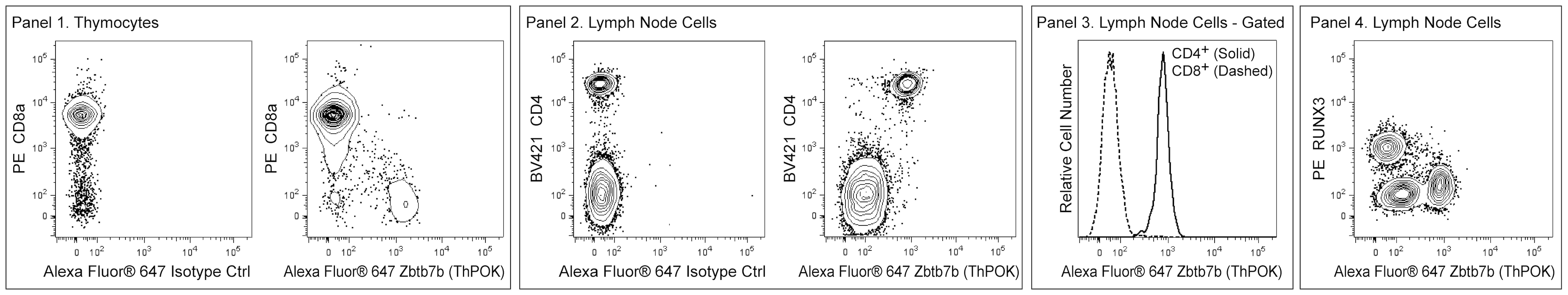
The T43-94 monoclonal antibody specifically recognizes mouse Zinc finger and BTB domain-containing protein 7B (Zbtb7b) which is also known as, T-helper-inducing POZ/Krueppel-like factor (Th-POK), Krueppel-related zinc finger protein cKrox (cKrox), or Zinc finger protein 67 (Zfp67). Zbtb7b is a Zn2+ finger-containing transcription factor that acts as a transcriptional repressor or activator in a promoter-dependent manner. Zbtb7b plays a role in thymic selection of T cells and is expressed in CD4+CD8+ thymocytes and mature CD4+CD8- thymocytes and T cells. It is not expressed by mature CD4-CD8+ thymocytes and T cells. It indirectly increases the expression of CD4 in developing T cells by antagonizing Runx-3-mediated CD4 repression. Zbtb7b expression has also been found in NKT and gamma/delta T cells. Zbtb7b plays a role in transcriptional repression of collagen gene expression as well.

565500 Rev. 1
Format Details
Down Arrow Up Arrow
Alexa Fluor™ 647
Alexa Fluor™ 647 Dye is part of the BD red family of dyes. This is a small organic fluorochrome with an excitation maximum (Ex Max) at 653-nm and an emission maximum (Em Max) at 669-nm. Alexa Fluor 647 is designed to be excited by the Red laser (627-640 nm) and detected using an optical filter centered near 520-nm (e.g., a 660/20 nm bandpass filter). Please ensure that your instrument’s configurations (lasers and optical filters) are appropriate for this dye.
Alexa Fluor™ 647
Red 627-640 nm
653 nm
669 nm
565500 Rev.1
Citations & References
Down Arrow Up Arrow

Development References (4)

  1. Kappes DJ. Expanding roles for ThPOK in thymic development. Immunol Rev. 2010; 238(1):182-194. (Biology). View Reference
  2. Luckey MA, Kimura MY, Waickman AT, Feigenbaum L, Singer A, Park JH. The transcription factor ThPOK suppresses Runx3 and imposes CD4(+) lineage fate by inducing the SOCS suppressors of cytokine signaling. Nat Immunol. Nat Immunol. 2014; 15(7):638-645. (Biology). View Reference
  3. Reis BS, Rogoz A, Costa-Pinto FA, Taniuchi I, Mucida D. Mutual expression of the transcription factors Runx3 and ThPOK regulates intestinal CD4(+) T cell immunity. Nat Immunol. 2013; 14(3):271-280. (Biology). View Reference
  4. Taniuchi I, Ellmeier W. Transcriptional and epigenetic regulation of CD4/CD8 lineage choice. Adv Immunol. 2011; 110:71-110. (Biology). View Reference
View All (4) View Less
565500 Rev. 1

Please refer to Support Documents for Quality Certificates


Global - Refer to manufacturer's instructions for use and related User Manuals and Technical data sheets before using this products as described


Comparisons, where applicable, are made against older BD Technology, manual methods or are general performance claims.  Comparisons are not made against non-BD technologies, unless otherwise noted.

For Research Use Only. Not for use in diagnostic or therapeutic procedures.