

Preparation And Storage
Recommended Assay Procedures
BD™ CompBeads can be used as surrogates to assess fluorescence spillover (Compensation). When fluorochrome conjugated antibodies are bound to CompBeads, they have spectral properties very similar to cells. However, for some fluorochromes there can be small differences in spectral emissions compared to cells, resulting in spillover values that differ when compared to biological controls. It is strongly recommended that when using a reagent for the first time, users compare the spillover on cell and CompBead to ensure that BD Comp beads are appropriate for your specific cellular application.
Product Notices
- This reagent has been pre-diluted for use at the recommended Volume per Test. We typically use 1 × 10^6 cells in a 100-µl experimental sample (a test).
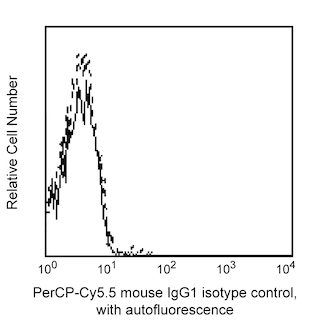
- An isotype control should be used at the same concentration as the antibody of interest.
- Caution: Sodium azide yields highly toxic hydrazoic acid under acidic conditions. Dilute azide compounds in running water before discarding to avoid accumulation of potentially explosive deposits in plumbing.
- Source of all serum proteins is from USDA inspected abattoirs located in the United States.
- PerCP-Cy5.5–labelled antibodies can be used with FITC- and R-PE–labelled reagents in single-laser flow cytometers with no significant spectral overlap of PerCP-Cy5.5, FITC, and R-PE fluorescence.
- Please observe the following precautions: Absorption of visible light can significantly alter the energy transfer occurring in any tandem fluorochrome conjugate; therefore, we recommend that special precautions be taken (such as wrapping vials, tubes, or racks in aluminum foil) to prevent exposure of conjugated reagents, including cells stained with those reagents, to room illumination.
- PerCP-Cy5.5 is optimized for use with a single argon ion laser emitting 488-nm light. Because of the broad absorption spectrum of the tandem fluorochrome, extra care must be taken when using dual-laser cytometers, which may directly excite both PerCP and Cy5.5™. We recommend the use of cross-beam compensation during data acquisition or software compensation during data analysis.
- For fluorochrome spectra and suitable instrument settings, please refer to our Multicolor Flow Cytometry web page at www.bdbiosciences.com/colors.
- Please refer to http://regdocs.bd.com to access safety data sheets (SDS).
- Cy is a trademark of Global Life Sciences Solutions Germany GmbH or an affiliate doing business as Cytiva.
- Please refer to www.bdbiosciences.com/us/s/resources for technical protocols.
Companion Products


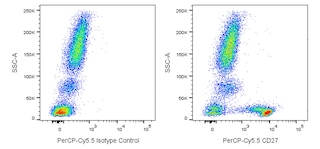
The O323 monoclonal antibody specifically recognizes CD27 which is also known as Tumor necrosis factor receptor superfamily member 7 (TNFRSF7), T14, Tp55, or S152. CD27 exists as a ~110-120 kDa disulfide-linked homodimer comprised of two single-pass type I transmembrane glycoproteins that are encoded by CD27 (CD27 molecule). CD27 is expressed on medullary thymocytes and T cells, with higher expression on activated T cells, and subsets of mature B cells and natural killer (NK) cells. A soluble 28-32 kDa form of CD27 is produced by lymphocytes upon cellular activation. Binding of the CD27 antigen, expressed on T cells, to its ligand, CD70 (CD27L), provides a costimulatory signal, leading to T cell proliferation, production of cytotoxic T cells, and enhanced production of cytokines. Binding of CD70 to CD27 expressed on B cells leads to B cell proliferation and the generation of plasma cells and immunoglobulin production. The CD27 antigen becomes hyperphosphorylated on serine residues upon activation of T cells. Signaling through the CD27 antigen activates NFκB and stress activated protein kinase (SAPK)/c Jun N terminal kinase (JNK).

Development References (5)
-
Björkström NK, Béziat V, Cichocki F, et al. CD8 T cells express randomly selected KIRs with distinct specificities compared with NK cells.. Blood. 2012; 120(17):3455-65. (Clone-specific: Flow cytometry). View Reference
-
Borst J, Hendriks J, Xiao Y. CD27 and CD70 in T cell and B cell activation.. Curr Opin Immunol. 2005; 17(3):275-81. (Biology). View Reference
-
Klein U, Rajewsky K, Küppers R. Human immunoglobulin (Ig)M+IgD+ peripheral blood B cells expressing the CD27 cell surface antigen carry somatically mutated variable region genes: CD27 as a general marker for somatically mutated (memory) B cells.. J Exp Med. 1998; 188(9):1679-89. (Biology). View Reference
-
Reiter C. T9. Cluster report: CD27. In: Knapp W. W. Knapp .. et al., ed. Leucocyte typing IV : white cell differentiation antigens. Oxford New York: Oxford University Press; 1989:350.
-
Zola H. Leukocyte and stromal cell molecules : the CD markers. Hoboken, N.J.: Wiley-Liss; 2007.
Please refer to Support Documents for Quality Certificates
Global - Refer to manufacturer's instructions for use and related User Manuals and Technical data sheets before using this products as described
Comparisons, where applicable, are made against older BD Technology, manual methods or are general performance claims. Comparisons are not made against non-BD technologies, unless otherwise noted.
For Research Use Only. Not for use in diagnostic or therapeutic procedures.
Refer to manufacturer's instructions for use and related User Manuals and Technical Data Sheets before using this product as described.
Comparisons, where applicable, are made against older BD technology, manual methods or are general performance claims. Comparisons are not made against non-BD technologies, unless otherwise noted.