
Preparation And Storage
Recommended Assay Procedures
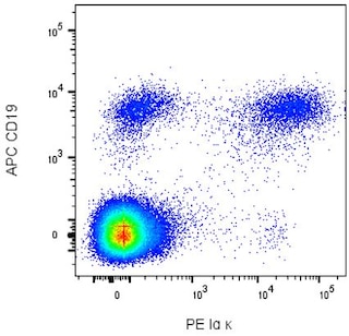
Some B lymphocytes and non-B leucocytes express Fc Receptors that can selectively bind various immunoglobulins from the plasma. As a result, these "cytophilic antibodies" can be detected on the cell surface using anti-Ig antibodies. Overnight incubation of peripheral blood mononuclear cells (PBMC) at 37ºC in complete tissue culture medium reportedly reduces the amount of these cytophilic antibodies observed on the PBMC. Costaining the PBMC with anti-CD20 or anti-CD19 and gating on the B lymphocytes eliminates detection of the cytophilic antibodies on the non-B cell populations, but a small proportion of the gated B lymphocytes may still express cytophilic antibodies.
BD™ CompBeads can be used as surrogates to assess fluorescence spillover (Compensation). When fluorochrome conjugated antibodies are bound to CompBeads, they have spectral properties very similar to cells. However, for some fluorochromes there can be small differences in spectral emissions compared to cells, resulting in spillover values that differ when compared to biological controls. It is strongly recommended that when using a reagent for the first time, users compare the spillover on cell and CompBead to ensure that BD Comp beads are appropriate for your specific cellular application.
Product Notices
- This reagent has been pre-diluted for use at the recommended Volume per Test. We typically use 1 × 10^6 cells in a 100-µl experimental sample (a test).
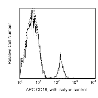
- An isotype control should be used at the same concentration as the antibody of interest.
- Caution: Sodium azide yields highly toxic hydrazoic acid under acidic conditions. Dilute azide compounds in running water before discarding to avoid accumulation of potentially explosive deposits in plumbing.
- Source of all serum proteins is from USDA inspected abattoirs located in the United States.
- For fluorochrome spectra and suitable instrument settings, please refer to our Multicolor Flow Cytometry web page at www.bdbiosciences.com/colors.
- Please refer to http://regdocs.bd.com to access safety data sheets (SDS).
- Please refer to www.bdbiosciences.com/us/s/resources for technical protocols.
Companion Products


.png?imwidth=320)
The Goat Anti-Human Ig λ Light Chain polyclonal antibody specifically recognizes human immunoglobulin lambda light chains (Igλ). Immunoglobulins are expressed on the surface of B lymphocytes and plasma cells, and they also circulate in the serum. The two light chains in an immunoglobulin are of the same type, either kappa (Igκ) or Igλ. Immunoglobulins bearing Igλ are expressed on a minority of normal B lymphocytes and on neoplastic cells of some leukemias, lymphomas, and plasmacytomas. In ELISA, the Goat Anti-Human Ig λ Light Chain polyclonal antibody binds to immunoglobulins bearing Igλ and not to immunoglobulins bearing Igκ. Cross-reactivity to Igλ of other species is possible.
The polyclonal antibody is derived from pooled antisera obtained from hyperimmunized goats, is purified by affinity chromatography on human Igλ, and is cross-adsorbed against human Igκ. It is provided as F(ab')2 fragments.

Development References (4)
-
Fukushima PI, Nguyen PK, O'Grady P, Stetler-Stevenson M. Flow cytometric analysis of kappa and lambda light chain expression in evaluation of specimens for B-cell neoplasia.. Cytometry. 1996; 26(4):243-52. (Methodology: Flow cytometry). View Reference
-
Kubagawa H, Gathings WE, Levitt D, Kearney JF, Cooper MD. Immunoglobulin isotype expression of normal pre-B cells as determined by immunofluorescence.. J Clin Immunol. 1982; 2(4):264-9. (Biology: Immunofluorescence). View Reference
-
Picker LJ, Weiss LM, Medeiros LJ, Wood GS, Warnke RA. Immunophenotypic criteria for the diagnosis of non-Hodgkin's lymphoma.. Am J Pathol. 1987; 128(1):181-201. (Biology: Immunohistochemistry). View Reference
-
Smith BR, Weinberg DS, Robert NJ, et al. Circulating monoclonal B lymphocytes in non-Hodgkin's lymphoma.. N Engl J Med. 1984; 311(23):1476-81. (Biology: Flow cytometry). View Reference
Please refer to Support Documents for Quality Certificates
Global - Refer to manufacturer's instructions for use and related User Manuals and Technical data sheets before using this products as described
Comparisons, where applicable, are made against older BD Technology, manual methods or are general performance claims. Comparisons are not made against non-BD technologies, unless otherwise noted.
For Research Use Only. Not for use in diagnostic or therapeutic procedures.
Refer to manufacturer's instructions for use and related User Manuals and Technical Data Sheets before using this product as described.
Comparisons, where applicable, are made against older BD technology, manual methods or are general performance claims. Comparisons are not made against non-BD technologies, unless otherwise noted.