Old Browser
Looks like you're visiting us from {countryName}.
Would you like to stay on the current country site or be switched to your country?


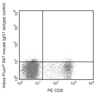
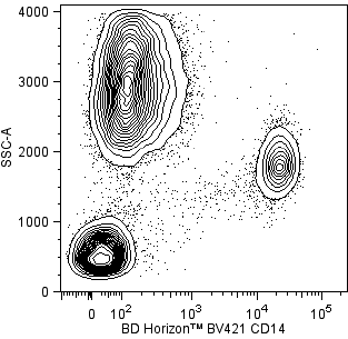
Flow cytometric analysis of Heme oxygenase 1 expression in human peripheral blood mononuclear cells. Peripheral blood mononuclear cells (PBMCs) were fixed and permeabilized with BD Cytofix/Cytoperm™ Fixation and Permeabilization Solution (Cat. No. 554722). The cells were then washed and stained in BD Perm/Wash™ Buffer (Cat. No. 554723) with BD Horizon™ BV421 Mouse Anti-Human CD14 antibody (Cat. No. 565283) and either Alexa Fluor® 647 Mouse IgG1, κ Isotype Control (Cat. No.557732; Left Plot) or Alexa Fluor® 647 Mouse Anti-Human Heme oxygenase 1 antibody (Cat. No. 566391; Right Plot). Two-color flow cytometric contour plots showing the correlated expression of Heme oxygenase 1 (or Ig Isotype control staining) versus CD14 were derived from gated events with the forward and side light-scatter characteristics of intact leucocytes. Flow cytometric analysis was performed using a BD LSRFortessa™ X-20 Cell Analyzer System. Data shown on this Technical Data Sheet are not lot specific.


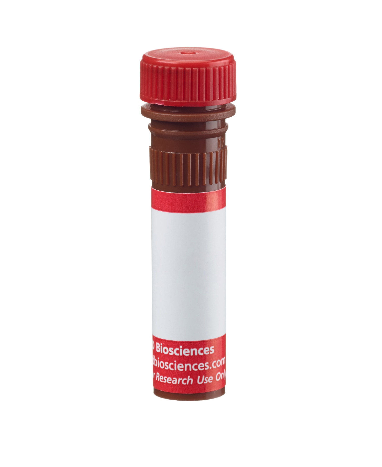
BD Pharmingen™ Alexa Fluor® 647 Mouse Anti-Heme Oxygenase 1

Regulatory Status Legend
Any use of products other than the permitted use without the express written authorization of Becton, Dickinson and Company is strictly prohibited.
Preparation And Storage
Product Notices
- This reagent has been pre-diluted for use at the recommended Volume per Test. We typically use 1 × 10^6 cells in a 100-µl experimental sample (a test).
- An isotype control should be used at the same concentration as the antibody of interest.
- Caution: Sodium azide yields highly toxic hydrazoic acid under acidic conditions. Dilute azide compounds in running water before discarding to avoid accumulation of potentially explosive deposits in plumbing.
- Source of all serum proteins is from USDA inspected abattoirs located in the United States.
- The Alexa Fluor®, Pacific Blue™, and Cascade Blue® dye antibody conjugates in this product are sold under license from Molecular Probes, Inc. for research use only, excluding use in combination with microarrays, or as analyte specific reagents. The Alexa Fluor® dyes (except for Alexa Fluor® 430), Pacific Blue™ dye, and Cascade Blue® dye are covered by pending and issued patents.
- Alexa Fluor® is a registered trademark of Molecular Probes, Inc., Eugene, OR.
- Alexa Fluor® 647 fluorochrome emission is collected at the same instrument settings as for allophycocyanin (APC).
- For fluorochrome spectra and suitable instrument settings, please refer to our Multicolor Flow Cytometry web page at www.bdbiosciences.com/colors.
- Please refer to www.bdbiosciences.com/us/s/resources for technical protocols.
Companion Products


The 23/Heme Oxygenase 1 monoclonal antibody specifically recognizes Heme oxygenase 1 (HO-1), also known as Heat Shock Protein 32 (HSP32). HO-1 is a 32 kDa monooxygenase encoded by HMOX1. Heme oxygenases 1 and 2 (HO-1, HO-2) cleave the heme molecule, resulting in the production of carbon monoxide (CO) and biliverdin. While HO-2 is constitutively expressed in tissues, HO-1 is rapidly induced by several stimuli such as lutathione depletion, hemin, heat shock, heavy metals, oxidative stress, oxidized LDL, anoxia, and endotoxic shock. HO-1 and its byproducts have potent antioxidant, antiproliferative, anti-inflammatory and anti-apoptotic effects in various cell types. In humans, HO-1 deficiency is associated with susceptibility to oxidative stress, increased inflammation and endothelial cell damage. In immune cells, HO-1 is required for the activation of interferon (IFN) regulatory factor 3 (IRF3) after Toll-like receptor 3 or 4 stimulation or viral infection. It is also upregulated on T cells upon TCR stimulation.
Development References (8)
-
Foresti R, Clark JE, Green CJ, Motterlini R. Thiol compounds interact with nitric oxide in regulating heme oxygenase-1 induction in endothelial cells. Involvement of superoxide and peroxynitrite anions. J Biol Chem. 1997; 272(29):18411-18417. (Biology). View Reference
-
Fürst R, Blumenthal SB, Kiemer AK, Zahler S, Vollmar AM. Nuclear factor-kappa B-independent anti-inflammatory action of salicylate in human endothelial cells: induction of heme oxygenase-1 by the c-jun N-terminal kinase/activator protein-1 pathway.. J Pharmacol Exp Ther. 2006; 318(1):389-94. (Clone-specific: Western blot). View Reference
-
Ishikawa K, Navab M, Leitinger N, Fogelman AM, Lusis AJ. Induction of heme oxygenase-1 inhibits the monocyte transmigration induced by mildly oxidized LDL. J Clin Invest. 1997; 100(5):1209-1216. (Biology). View Reference
-
McAllister SC, Hansen SG, Ruhl RA, et al. Kaposi sarcoma-associated herpesvirus (KSHV) induces heme oxygenase-1 expression and activity in KSHV-infected endothelial cells.. Blood. 2004; 103(9):3465-73. (Clone-specific: Immunofluorescence). View Reference
-
Suzuki M, Ishizaka N, Tsukamoto K. Pressurization facilitates adenovirus-mediated gene transfer into vein graft. FEBS Lett. 2000; 470(3):370-374. (Clone-specific: Western blot). View Reference
-
Wang X, Mazurkiewicz M, Hillert EK, et al. The proteasome deubiquitinase inhibitor VLX1570 shows selectivity for ubiquitin-specific protease-14 and induces apoptosis of multiple myeloma cells.. Sci Rep. 2016; 6:26979. (Clone-specific: Western blot). View Reference
-
Yoshida T, Biro P, Cohen T, Muller RM, Shibahara S. Human heme oxygenase cDNA and induction of its mRNA by hemin. Eur J Biochem. 1988; 171(3):457-461. (Biology). View Reference
-
Zhong H, Bao W, Friedman D, Yazdanbakhsh K. Hemin controls T cell polarization in sickle cell alloimmunization.. J Immunol. 2014; 193(1):102-10. (Clone-specific: Flow cytometry). View Reference
Please refer to Support Documents for Quality Certificates
Global - Refer to manufacturer's instructions for use and related User Manuals and Technical data sheets before using this products as described
Comparisons, where applicable, are made against older BD Technology, manual methods or are general performance claims. Comparisons are not made against non-BD technologies, unless otherwise noted.
For Research Use Only. Not for use in diagnostic or therapeutic procedures.
Refer to manufacturer's instructions for use and related User Manuals and Technical Data Sheets before using this product as described.
Comparisons, where applicable, are made against older BD technology, manual methods or are general performance claims. Comparisons are not made against non-BD technologies, unless otherwise noted.
Report a Site Issue
This form is intended to help us improve our website experience. For other support, please visit our Contact Us page.