Skip to main content Skip to navigation
PE Mouse Anti-ID3
PE Mouse Anti-ID3
Flow cytometric analysis of ID3 expression in human Ramos cells. Cells from the human Ramos (Burkitt's lymphoma, ATCC CRL-1596) cell line were fixed and permeabilized with the BD Pharmingen™ Transcription Factor Buffer Set (Cat. No. 562574/562725). The cells were then stained with either PE Mouse IgG1, κ Isotype Control (Cat No. 554680; dotted line histogram) or PE Mouse Anti-ID3 antibody (Cat. No. 564564; solid line histogram). The fluorescence histograms were derived from events with forward and side light-scatter characteristics of intact Ramos cells. Flow cytometric analysis was performed using a BD FACSCanto™ II Flow Cytometer System.
PE Mouse Anti-ID3
Flow cytometric analysis of ID3 expression in mouse thymocytes. Mouse thymocytes were stimulated for 3 hours with Phorbol 12-Myristate 13-Acetate (10 ng/ml). The cells were fixed and permeabilized with the BD Pharmingen™ Transcription Factor Buffer Set and preincubated with Purified Rat Anti-Mouse CD16/CD32 antibody (Mouse BD Fc Block™) (Cat. No. 553141/553142). The cells were then stained with either PE Mouse IgG1 Isotype Control (dotted line histogram) or PE Mouse Anti-ID3 antibody (solid line histogram). The mouse thymocytes were analyzed by flow cytometry as described for the human Ramos cells.
Flow cytometric analysis of ID3 expression in human Ramos cells. Cells from the human Ramos (Burkitt's lymphoma, ATCC CRL-1596) cell line were fixed and permeabilized with the BD Pharmingen™ Transcription Factor Buffer Set (Cat. No. 562574/562725). The cells were then stained with either PE Mouse IgG1, κ Isotype Control (Cat No. 554680; dotted line histogram) or PE Mouse Anti-ID3 antibody (Cat. No. 564564; solid line histogram). The fluorescence histograms were derived from events with forward and side light-scatter characteristics of intact Ramos cells. Flow cytometric analysis was performed using a BD FACSCanto™ II Flow Cytometer System.
Flow cytometric analysis of ID3 expression in mouse thymocytes. Mouse thymocytes were stimulated for 3 hours with Phorbol 12-Myristate 13-Acetate (10 ng/ml). The cells were fixed and permeabilized with the BD Pharmingen™ Transcription Factor Buffer Set and preincubated with Purified Rat Anti-Mouse CD16/CD32 antibody (Mouse BD Fc Block™) (Cat. No. 553141/553142). The cells were then stained with either PE Mouse IgG1 Isotype Control (dotted line histogram) or PE Mouse Anti-ID3 antibody (solid line histogram). The mouse thymocytes were analyzed by flow cytometry as described for the human Ramos cells.
Product Details
Down Arrow Up Arrow


BD Pharmingen™
ID-3; Inhibitor of DNA binding 3; 1R21; BHLHB25; HEIR1; HEIR-1
Human (QC Testing), Mouse (Tested in Development)
Mouse IgG1, κ
Human ID3 Recombinant Protein
Intracellular staining (flow cytometry) (Routinely Tested)
0.2 mg/ml
AB_2738846
Aqueous buffered solution containing ≤0.09% sodium azide.
RUO


Preparation And Storage

Store undiluted at 4°C and protected from prolonged exposure to light. Do not freeze. The monoclonal antibody was purified from tissue culture supernatant or ascites by affinity chromatography. The antibody was conjugated with R-PE under optimum conditions, and unconjugated antibody and free PE were removed.

Product Notices

  1. Since applications vary, each investigator should titrate the reagent to obtain optimal results.
  2. Please refer to www.bdbiosciences.com/us/s/resources for technical protocols.
  3. An isotype control should be used at the same concentration as the antibody of interest.
  4. Caution: Sodium azide yields highly toxic hydrazoic acid under acidic conditions. Dilute azide compounds in running water before discarding to avoid accumulation of potentially explosive deposits in plumbing.
  5. For fluorochrome spectra and suitable instrument settings, please refer to our Multicolor Flow Cytometry web page at www.bdbiosciences.com/colors.
564564 Rev. 1
Antibody Details
Down Arrow Up Arrow
S30-778

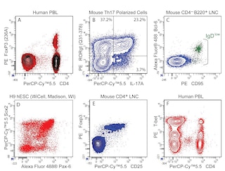
The S30-778 monoclonal antibody specifically binds to Inhibitor of DNA binding 3 (ID3). ID3 is one of four ID proteins (ID1-4) that have been characterized in humans. It is also known as Class B basic helix-loop-helix protein 25 (BHLHB25), Helix-loop-helix protein HEIR-1, and ID-like protein inhibitor HLH 1R21. ID3 is a Helix-Loop-Helix (HLH) protein that can form heterodimers with other HLH transcription factors such as MyoD, myogenin, E12 and E47. These latter transcription factors normally regulate the development, differentiation and functions of a variety of cell types including muscle cells, and subsets of T and B cells. Because it lacks a basic DNA-binding domain, ID3 can inhibit the DNA binding and function of these other transcription factors it complexes with. Abnormal ID3 expression has been associated with certain cancers. The S30-778.8.66 antibody crossreacts with mouse Id3.

564564 Rev. 1
Format Details
Down Arrow Up Arrow
PE
R-Phycoerythrin (PE), is part of the BD family of Phycobiliprotein dyes. This fluorochrome is a multimeric fluorescent phycobiliprotein with excitation maximum (Ex Max) of 496 nm and 566 nm and an emission maximum (Em Max) at 576 nm. PE is designed to be excited by the Blue (488 nm), Green (532 nm) and Yellow-Green (561 nm) lasers and detected using an optical filter centered near 575 nm (e.g., a 575/26-nm bandpass filter). As PE is excited by multiple lasers, this can result in cross-laser excitation and fluorescence spillover on instruments with various combinations of Blue, Green, and Yellow-Green lasers. Please ensure that your instrument’s configurations (lasers and optical filters) are appropriate for this dye.
altImg
PE
Yellow-Green 488 nm, 532 nm, 561 nm
496 nm, 566 nm
576 nm
564564 Rev.1
Citations & References
Down Arrow Up Arrow

Development References (6)

  1. Ishiguro A, Spirin K, Shiohara M, et al. Expression of Id2 and Id3 mRNA in human lymphocytes. Leuk Res. 1995; 19(12):989-996. (Biology). View Reference
  2. Lasorella A, Uo T, Iavarone A. Id proteins at the cross-road of development and cancer. Oncogene. 201; 20(58):8326-8333. (Biology). View Reference
  3. Li J1, Maruyama T, Zhang P, et al. Mutation of inhibitory helix-loop-helix protein Id3 causes γδ T-cell lymphoma in mice. Blood. 2010; 116(25):5615-5621. (Biology). View Reference
  4. Maruyama T1, Li J, Vaque JP, et al. Control of the differentiation of regulatory T cells and T(H)17 cells by the DNA-binding inhibitor Id3. Nat Immunol. 2011; 12(1):86-95. (Biology). View Reference
  5. Miyazaki M1, Rivera RR, Miyazaki K, Lin YC, Agata Y, Murre C. The opposing roles of the transcription factor E2A and its antagonist Id3 that orchestrate and enforce the naive fate of T cells. Nat Immunol. 2011; 12(10):992-1001. (Biology). View Reference
  6. Yokota Y. Id and development. Oncogene. 2001; 20(58):8290-8298. (Biology). View Reference
View All (6) View Less
564564 Rev. 1

Please refer to Support Documents for Quality Certificates


Global - Refer to manufacturer's instructions for use and related User Manuals and Technical data sheets before using this products as described


Comparisons, where applicable, are made against older BD Technology, manual methods or are general performance claims.  Comparisons are not made against non-BD technologies, unless otherwise noted.

For Research Use Only. Not for use in diagnostic or therapeutic procedures.