BD Pharmingen™ PE-Cy™5 Streptavidin
(RUO)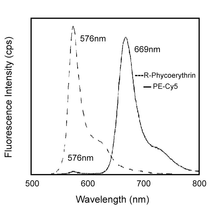
Preparation And Storage
Recommended Assay Procedures
PE-Cy5 tandem fluorochromes have been reported to bind some classes of human macrophages and granulocytes via Fc receptors, and PE has been reported to bind to mouse B lymphocytes via Fc receptors. Preincubation of mouse leukocytes with Purified Rat Anti-Mouse CD16/CD32 (Mouse BD Fc Block™) (Cat. No. 553141/553142) can reduce the non-specific binding of PE-Cy5-conjugated reagents to mouse B cells. For analysis of human peripheral blood, gating on specific cell populations based upon forward and light scatter, plus lineage markers, can help to overcome this limitation.
Furthermore, we have observed a distinct type of interaction between PE-Cy5 tandem fluorochromes and the splenocytes of SJL, NOD, and MRL mice: B lymphocytes and some other leukocyte subsets are brightly stained, and Mouse BD Fc Block™ has no significant effect. Therefore, PE-Cy5-conjugated reagents should not be used to stain leukocytes of SJL, NOD, or MRL mice. Reagents conjugated to PE, PerCP, PerCP-Cy5.5, Allophycocyanin (APC), and APC-Cy7 tandem fluorochrome can be used on leukocytes from these mouse strains.
Product Notices
- Since applications vary, each investigator should titrate the reagent to obtain optimal results.
- Caution: Sodium azide yields highly toxic hydrazoic acid under acidic conditions. Dilute azide compounds in running water before discarding to avoid accumulation of potentially explosive deposits in plumbing.
- For fluorochrome spectra and suitable instrument settings, please refer to our Multicolor Flow Cytometry web page at www.bdbiosciences.com/colors.
- PE-Cy5 is a tandem fluorochrome composed of R-phycoerythrin (PE), which is excited by the 488 nm light of an Argon ion laser and serves as an energy donor, coupled to the cyanine dye Cy5, which acts as an energy acceptor and fluoresces at 670 nm. BD Biosciences Pharmingen has maximized the fluorochrome energy transfer in PE-Cy5, thus maximizing its fluorescence emission intensity, minimizing residual emission from PE, and minimizing lot-to-lot variation.
- PE-Cy5 is optimized for use with a single argon ion laser emitting 488-nm light. Because of the broad absorption spectrum of the PE-Cy5 tandem fluorochrome, extra care must be taken when using dual-laser cytometers which may directly excite both PE and Cy5™.
- Please observe the following precautions: Absorption of visible light can significantly alter the energy transfer occurring in any tandem fluorochrome conjugate; therefore, we recommend that special precautions be taken (such as wrapping vials, tubes, or racks in aluminum foil) to prevent exposure of conjugated reagents, including cells stained with those reagents, to room illumination.
- PE-Cy5 tandem fluorochromes have been reported to bind some classes of human macrophages and granulocytes via Fc receptors, and PE has been reported to bind to mouse B lymphocytes via Fc receptors. Preincubation of mouse leukocytes with Mouse BD Fc Block™ purified anti-mouse CD16/CD32 mAb 2.4G2 can reduce the non-specific binding of PE-Cy5-conjugated reagents to mouse B cells. However, PE-Cy5 conjugated reagents should not be used to stain splenocytes of SJL, NOD, and MRL mice as B lymphocytes and/or other leukocytes have been reported to non-specifically stain regardless of the use of Mouse BD Fc Block™ (the CD72c complex has been implicated for PE-Cy5 binding in these strains). Reagents conjugated to PE, PerCP, PerCP-Cy5.5, APC, and APC-Cy7 tandem fluorochrome can be used on leukocytes from these mouse strains.
- Cy is a trademark of GE Healthcare.
- Please refer to http://regdocs.bd.com to access safety data sheets (SDS).
- Please refer to www.bdbiosciences.com/us/s/resources for technical protocols.
Companion Products

SAv-PE-Cy5 is a useful second-step reagent for the indirect immunofluorescent staining of cells in combination with biotinylated primary antibodies for flow cytometric analysis. Excitation at 488-nm light leads to a fluorescence emission maximum of ~669 nm.
Development References (4)
-
Stewart CC, Stewart SJ. Immunological monitoring utilizing novel probes. Ann N Y Acad Sci. 1993; 677:94-112. (Biology).
-
Takizawa F, Kinet JP, Adamczewski M. Binding of phycoerythrin and its conjugates to murine low affinity receptors for immunoglobulin G. J Immunol Methods. 1993; 162(2):269-272. (Biology). View Reference
-
Waggoner AS, Ernst LA, Chen CH, Rechtenwald DJ. PE-CY5. A new fluorescent antibody label for three-color flow cytometry with a single laser. Ann N Y Acad Sci. 1993; 677:185-193. (Biology). View Reference
-
van Vugt MJ, van den Herik-Oudijk IE, van de Winkle JG. Binding of PE-CY5 conjugates to the human high-affinity receptor for IgG (CD64). Blood. 1996; 88(6):2358-2361. (Biology). View Reference
Please refer to Support Documents for Quality Certificates
Global - Refer to manufacturer's instructions for use and related User Manuals and Technical data sheets before using this products as described
Comparisons, where applicable, are made against older BD Technology, manual methods or are general performance claims. Comparisons are not made against non-BD technologies, unless otherwise noted.
For Research Use Only. Not for use in diagnostic or therapeutic procedures.