Skip to main content Skip to navigation
Alexa Fluor® 647 Mouse Anti-Cardiac Troponin I
Analysis of Cardiac Troponin I (cTnI) in human embryonic stem cell (hESC)-derived cardiomyocytes. hESC-derived cardiomyocytes (Evans lab, UCSD) were disassociated and fixed in BD Cytofix™ Fixation Buffer (Cat. No. 554655), permeabilized with BD Phosflow™ Perm/Wash Buffer I (Cat. No. 557885) and stained with PE Mouse Anti-Myosin Heavy Chain (Cat. No. 564408) and Alexa Fluor® 647 Mouse Anti-Cardiac Troponin I. The cells were first gated on light scatter properties and then analyzed for Myosin Heavy Chain and cTnI expression.  Flow cytometry was performed on a BD LSRFortessa™ Cell Analyzer System.
Alexa Fluor® 647 Mouse Anti-Cardiac Troponin I
Analysis of Cardiac Troponin I (cTnI) in differentiated C2C12 mouse myoblast cell line. C2C12 cells (ATCC CRL-1772) were differentiated for 5 days and then disassociated and fixed in BD Cytofix™ Fixation buffer (Cat. No. 554655), permeabilized with BD Phosflow™ Perm/Wash buffer I (Cat. No. 557885) and stained with Alexa Fluor® 647 Mouse Anti-Cardiac Troponin I. The cells were first gated on light scatter properties and then analyzed for cTnI expression.  Flow cytometry was performed on a BD LSRFortessa™ Cell Analyzer System.
Analysis of Cardiac Troponin I (cTnI) in human embryonic stem cell (hESC)-derived cardiomyocytes. hESC-derived cardiomyocytes (Evans lab, UCSD) were disassociated and fixed in BD Cytofix™ Fixation Buffer (Cat. No. 554655), permeabilized with BD Phosflow™ Perm/Wash Buffer I (Cat. No. 557885) and stained with PE Mouse Anti-Myosin Heavy Chain (Cat. No. 564408) and Alexa Fluor® 647 Mouse Anti-Cardiac Troponin I. The cells were first gated on light scatter properties and then analyzed for Myosin Heavy Chain and cTnI expression.  Flow cytometry was performed on a BD LSRFortessa™ Cell Analyzer System.
Analysis of Cardiac Troponin I (cTnI) in differentiated C2C12 mouse myoblast cell line. C2C12 cells (ATCC CRL-1772) were differentiated for 5 days and then disassociated and fixed in BD Cytofix™ Fixation buffer (Cat. No. 554655), permeabilized with BD Phosflow™ Perm/Wash buffer I (Cat. No. 557885) and stained with Alexa Fluor® 647 Mouse Anti-Cardiac Troponin I. The cells were first gated on light scatter properties and then analyzed for cTnI expression.  Flow cytometry was performed on a BD LSRFortessa™ Cell Analyzer System.
Product Details
Down Arrow Up Arrow


BD Pharmingen™
cTnI, TNNI3, TNNC1
Mouse (QC Testing), Human (Tested in Development)
Mouse BALB/c IgG2b, κ
Human
Intracellular staining (flow cytometry) (Routinely Tested)
5 µl/test
AB_2738796
Aqueous buffered solution containing BSA, protein stabilizer, and ≤0.09% sodium azide.
RUO


Preparation And Storage

Store undiluted at 4°C and protected from prolonged exposure to light. Do not freeze. The monoclonal antibody was purified from tissue culture supernatant or ascites by affinity chromatography. The antibody was conjugated to Alexa Fluor® 647 under optimum conditions, and unreacted Alexa Fluor® 647 was removed.

Product Notices

  1. This reagent has been pre-diluted for use at the recommended Volume per Test. We typically use 1 × 10^6 cells in a 100-µl experimental sample (a test).
  2. An isotype control should be used at the same concentration as the antibody of interest.
  3. Alexa Fluor® is a registered trademark of Molecular Probes, Inc., Eugene, OR.
  4. Alexa Fluor® 647 fluorochrome emission is collected at the same instrument settings as for allophycocyanin (APC).
  5. The Alexa Fluor®, Pacific Blue™, and Cascade Blue® dye antibody conjugates in this product are sold under license from Molecular Probes, Inc. for research use only, excluding use in combination with microarrays, or as analyte specific reagents. The Alexa Fluor® dyes (except for Alexa Fluor® 430), Pacific Blue™ dye, and Cascade Blue® dye are covered by pending and issued patents.
  6. Source of all serum proteins is from USDA inspected abattoirs located in the United States.
  7. Caution: Sodium azide yields highly toxic hydrazoic acid under acidic conditions. Dilute azide compounds in running water before discarding to avoid accumulation of potentially explosive deposits in plumbing.
  8. For fluorochrome spectra and suitable instrument settings, please refer to our Multicolor Flow Cytometry web page at www.bdbiosciences.com/colors.
  9. Please refer to www.bdbiosciences.com/us/s/resources for technical protocols.
564409 Rev. 1
Antibody Details
Down Arrow Up Arrow
C5

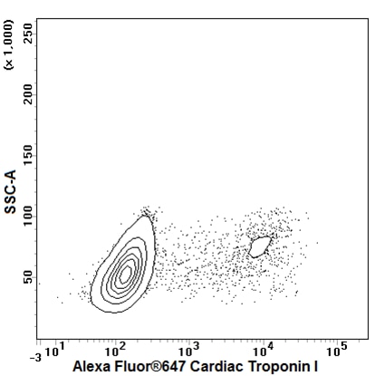
The C5 monoclonal antibody specifically binds to Cardiac Troponin I (cTnI). cTnI is a ~30-kDa protein that is also known as TNNI3 and TNNC1. Troponin I is the inhibitory subunit of Troponin, which also contains the subunits Troponin C and Troponin T. Troponin aids the Ca[2+]-dependent interaction of actin and myosin, which are the main contracile components in muscle cells. cTnI is specifically expressed in cardiac tissue, while skeletal troponin I is expressed in skeletal muscle in slow and fast isoforms. Hereditary cardiomyopathy is associated with mutations in cTnI. During product development, the purified C5 monoclonal antibody detected cTnI by western blot analysis of cellular lysates and flow cytometric analysis of fixed and permeabilized cells.

564409 Rev. 1
Format Details
Down Arrow Up Arrow
Alexa Fluor™ 647
Alexa Fluor™ 647 Dye is part of the BD red family of dyes. This is a small organic fluorochrome with an excitation maximum (Ex Max) at 653-nm and an emission maximum (Em Max) at 669-nm. Alexa Fluor 647 is designed to be excited by the Red laser (627-640 nm) and detected using an optical filter centered near 520-nm (e.g., a 660/20 nm bandpass filter). Please ensure that your instrument’s configurations (lasers and optical filters) are appropriate for this dye.
Alexa Fluor™ 647
Red 627-640 nm
653 nm
669 nm
564409 Rev.1
Citations & References
Down Arrow Up Arrow

Development References (5)

  1. Bhavsar PK, Brand NJ, Yacoub MH, Barton PJ. Isolation and characterization of the human cardiac troponin I gene (TNNI3). Genomics. 1996; 35(1):11-23. (Biology). View Reference
  2. Dubois NC, Craft AM, Sharma P, et al. SIRPA is a specific cell-surface marker for isolating cardiomyocytes derived from human pluripotent stem cells. Nat Biotechnol. 2011; 29:1011-1018. (Methodology). View Reference
  3. Katrukha AG, Bereznikova AV, Esakova TV, Filatov VL, Bulargina TV, Gusev NB. A new method of human cardiac troponin I and troponin T purification. 1995; 36(1):195-202. (Clone-specific). View Reference
  4. Kehat I, Kenyagin-Karsenti D, Snir M, et al. Human embryonic stem cells can differentiate into myocytes with structural and functional properties of cardiomyocytes. J Clin Invest. 2001; 108(3):407-414. (Biology). View Reference
  5. Norström A, Akesson K, Hardarson T, Hamberger L, Björquist P, Sartipy P. Molecular and pharmacological properties of human embryonic stem cell-derived cardiomyocytes. Exp Biol Med (Maywood). 2006; 231(11):1753-1762. (Biology). View Reference
View All (5) View Less
564409 Rev. 1

Please refer to Support Documents for Quality Certificates


Global - Refer to manufacturer's instructions for use and related User Manuals and Technical data sheets before using this products as described


Comparisons, where applicable, are made against older BD Technology, manual methods or are general performance claims.  Comparisons are not made against non-BD technologies, unless otherwise noted.

For Research Use Only. Not for use in diagnostic or therapeutic procedures.