Skip to main content Skip to navigation
PE Mouse Anti-Human TRAIL-R2 (CD262)
Flow cytometric analysis of TRAIL-R2 (CD262) expression on U937 cells. Cells from the human U937 (Histiocytic lymphoma, ATCC CRL-1593) cell line were stained with either PE Mouse IgG1, κ Isotype Control (Cat. No. 554680; dashed line histogram) or PE Mouse Anti-Human CD262 (TRAIL-R2) antibody (Cat. No. 565499).  The fluorescence histogram showing TRAIL-R2 (CD262) expression [or Ig Isotype control staining] was derived from gated events with the forward and side light-scatter characteristics of viable cells. Flow cytometric analysis was performed using a BD FACSCanto™ II Flow Cytometer System.
Product Details
Down Arrow Up Arrow


BD Pharmingen™
TNFRSF10B, DR5, KILLER, TRAILR2, TRICK2, ZTNFR9
Human (QC Testing)
Mouse BALB/c IgG1, κ
Human DR5/TRAIL-R2 Recombinant Protein
Flow cytometry (Routinely Tested)
5 µl/test
AB_2732871
Aqueous buffered solution containing BSA, protein stabilizer, and ≤0.09% sodium azide.
RUO


Preparation And Storage

Store undiluted at 4°C and protected from prolonged exposure to light. Do not freeze. The monoclonal antibody was purified from tissue culture supernatant or ascites by affinity chromatography. The antibody was conjugated with R-PE under optimum conditions, and unconjugated antibody and free PE were removed.

Product Notices

  1. This reagent has been pre-diluted for use at the recommended Volume per Test. We typically use 1 × 10^6 cells in a 100-µl experimental sample (a test).
  2. Please refer to www.bdbiosciences.com/us/s/resources for technical protocols.
  3. Caution: Sodium azide yields highly toxic hydrazoic acid under acidic conditions. Dilute azide compounds in running water before discarding to avoid accumulation of potentially explosive deposits in plumbing.
  4. For fluorochrome spectra and suitable instrument settings, please refer to our Multicolor Flow Cytometry web page at www.bdbiosciences.com/colors.
  5. Source of all serum proteins is from USDA inspected abattoirs located in the United States.
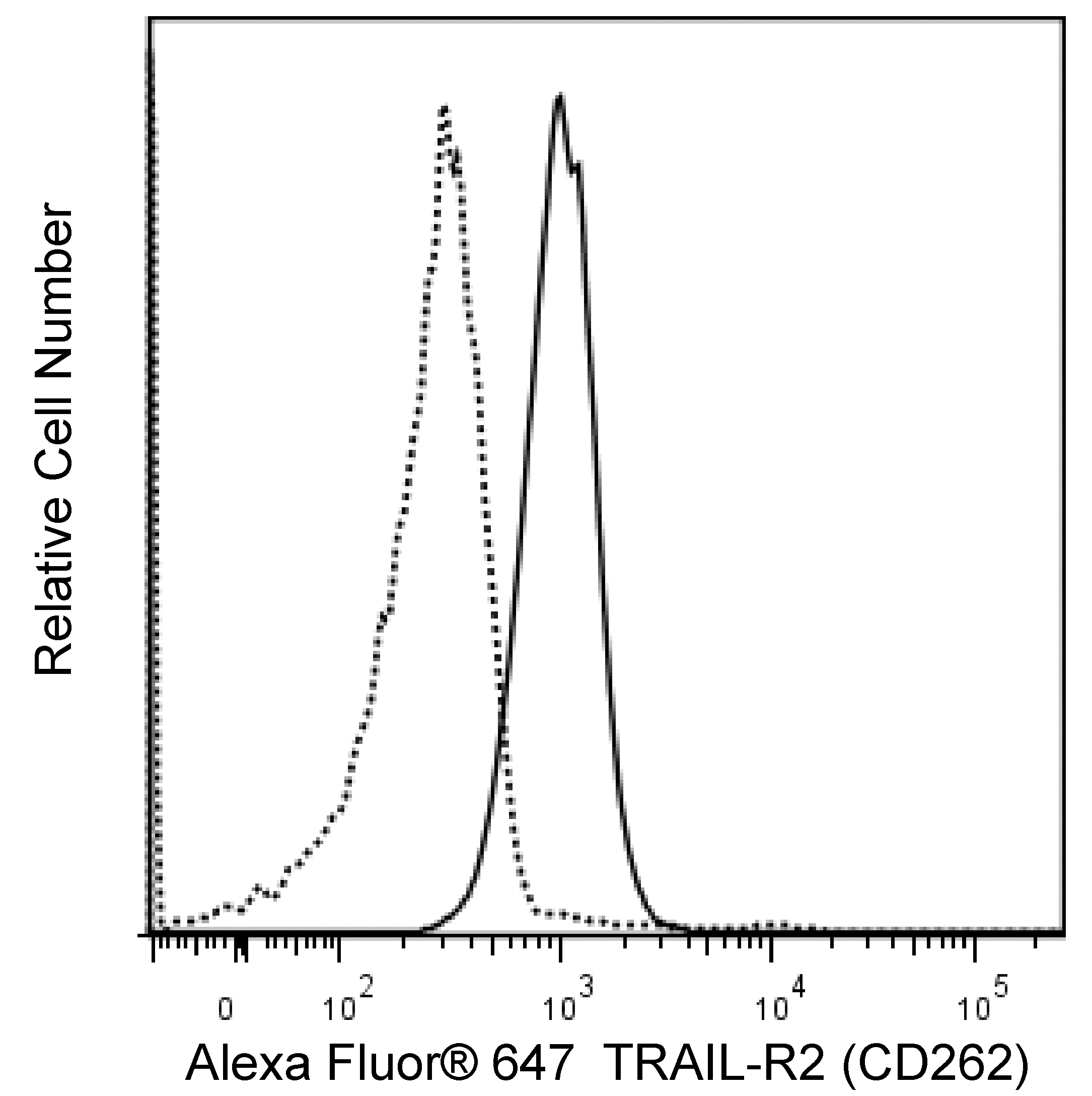
  6. An isotype control should be used at the same concentration as the antibody of interest.
565499 Rev. 1
Antibody Details
Down Arrow Up Arrow
YM366

The YM366 monoclonal antibody specifically binds to TNF-related apoptosis-inducing ligand receptor 2 (TRAIL Receptor 2, TRAIL-R2) which is also known as, CD262, Death receptor 5 (DR5), TRICK2, or KILLER. CD262 is a type I transmembrane protein that is encoded by TNFRSF10B (Tumor necrosis factor receptor superfamily, member 10b). CD262 forms a homotrimeric receptor complex that can bind homotrimeric TRAIL (CD253/APO-2 Ligand) and transduce apoptotic signals intracellularly through its cytoplasmic death domain (DD). CD262 is differentially expressed on cells from a wide variety of normal tissues and tumors. CD262 expression is upregulated by Interferon-α (IFN-α).

565499 Rev. 1
Format Details
Down Arrow Up Arrow
PE
R-Phycoerythrin (PE), is part of the BD family of Phycobiliprotein dyes. This fluorochrome is a multimeric fluorescent phycobiliprotein with excitation maximum (Ex Max) of 496 nm and 566 nm and an emission maximum (Em Max) at 576 nm. PE is designed to be excited by the Blue (488 nm), Green (532 nm) and Yellow-Green (561 nm) lasers and detected using an optical filter centered near 575 nm (e.g., a 575/26-nm bandpass filter). As PE is excited by multiple lasers, this can result in cross-laser excitation and fluorescence spillover on instruments with various combinations of Blue, Green, and Yellow-Green lasers. Please ensure that your instrument’s configurations (lasers and optical filters) are appropriate for this dye.
altImg
PE
Yellow-Green 488 nm, 532 nm, 561 nm
496 nm, 566 nm
576 nm
565499 Rev.1
Citations & References
Down Arrow Up Arrow

Development References (2)

  1. Liu F, Si Y, Liu G, Li S, Zhang J, Ma Y. The tetravalent anti-DR5 antibody without cross-linking direct induces apoptosis of cancer cells. Biomed Res Int. 2015; 70(0):41-45. (Clone-specific: Flow cytometry). View Reference
  2. Wang J, Lin Z, Qiao CX, et al. Characterization of a novel anti-DR5 monoclonal antibody WD1 with the potential to induce tumor cell apoptosis. Cell Mol Immunol. 2008; 5(1):55-60. (Clone-specific: Western blot). View Reference
565499 Rev. 1

Please refer to Support Documents for Quality Certificates


Global - Refer to manufacturer's instructions for use and related User Manuals and Technical data sheets before using this products as described


Comparisons, where applicable, are made against older BD Technology, manual methods or are general performance claims.  Comparisons are not made against non-BD technologies, unless otherwise noted.

For Research Use Only. Not for use in diagnostic or therapeutic procedures.