Skip to main content Skip to navigation
PE Mouse Anti-Human CD1c
Two-color flow cytometric analysis of CD1c expression on human peripheral blood lymphocytes. Human whole blood was treated with BD Pharm Lyse™ (Cat. No. 555899) to lyse erythrocytes. The leucocytes were stained with BD Horizon™ V450 Mouse Anti-Human CD19 antibody (Cat. No. 560353/560354) and either PE Mouse IgG1, κ Isotype Control (Cat. No. 554680; Left Panel) or PE Mouse Anti-Human CD1c (Cat. No. 564900; Right Panel). Two-color contour plots showing the correlated expression of CD19 versus CD1c  (or Ig Isotype control staining) were derived from gated events with the forward and side light-scatter characteristics of viable lymphocytes. Flow cytometric analysis was performed using a BD LSRFortessa™ Cell Analyzer System.
Product Details
Down Arrow Up Arrow


BD Pharmingen™
CD1; R7; M241; BDCA1
Human (QC Testing)
Mouse IgG1, κ
GM-CSF- and IL-4-activated Human Monocytes
Flow cytometry (Routinely Tested)
5 µl
911
AB_2739006
Aqueous buffered solution containing BSA and ≤0.09% sodium azide.
RUO


Preparation And Storage

Store undiluted at 4°C and protected from prolonged exposure to light. Do not freeze. The monoclonal antibody was purified from tissue culture supernatant or ascites by affinity chromatography. The antibody was conjugated with R-PE under optimum conditions, and unconjugated antibody and free PE were removed.

Product Notices

  1. This reagent has been pre-diluted for use at the recommended Volume per Test. We typically use 1 × 10^6 cells in a 100-µl experimental sample (a test).
  2. An isotype control should be used at the same concentration as the antibody of interest.
  3. Caution: Sodium azide yields highly toxic hydrazoic acid under acidic conditions. Dilute azide compounds in running water before discarding to avoid accumulation of potentially explosive deposits in plumbing.
  4. For fluorochrome spectra and suitable instrument settings, please refer to our Multicolor Flow Cytometry web page at www.bdbiosciences.com/colors.
  5. Source of all serum proteins is from USDA inspected abattoirs located in the United States.
  6. Please refer to www.bdbiosciences.com/us/s/resources for technical protocols.
564900 Rev. 2
Antibody Details
Down Arrow Up Arrow
F10/21A3

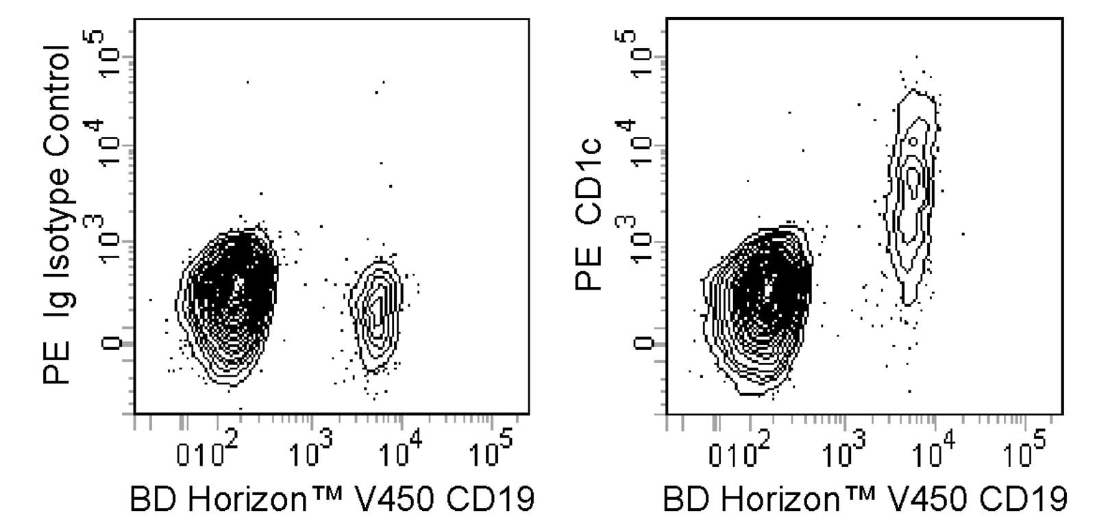
The F10/21A3 monoclonal antibody specifically binds to CD1c. The CD1 family of transmembrane glycoproteins are structurally related to the classical major histocompatibility complex (MHC) proteins. CD1c is a type I transmembrane glycoprotein that forms heterodimers with beta-2-microglobulin. CD1c presents lipids and glycolipids of self or microbial origin to T cells. CD1c is expressed by Langerhans cells, dendritic cells, monocytes, cortical thymocytes, T cells, and some B cells.

564900 Rev. 2
Format Details
Down Arrow Up Arrow
PE
R-Phycoerythrin (PE), is part of the BD family of Phycobiliprotein dyes. This fluorochrome is a multimeric fluorescent phycobiliprotein with excitation maximum (Ex Max) of 496 nm and 566 nm and an emission maximum (Em Max) at 576 nm. PE is designed to be excited by the Blue (488 nm), Green (532 nm) and Yellow-Green (561 nm) lasers and detected using an optical filter centered near 575 nm (e.g., a 575/26-nm bandpass filter). As PE is excited by multiple lasers, this can result in cross-laser excitation and fluorescence spillover on instruments with various combinations of Blue, Green, and Yellow-Green lasers. Please ensure that your instrument’s configurations (lasers and optical filters) are appropriate for this dye.
altImg
PE
Yellow-Green 488 nm, 532 nm, 561 nm
496 nm, 566 nm
576 nm
564900 Rev.2
Citations & References
Down Arrow Up Arrow

Development References (3)

  1. Delia D, Cattoretti G, Polli N, et al. CD1c but neither CD1a nor CD1b molecules are expressed on normal, activated, and malignant human B cells: identification of a new B-cell subset. Blood. 1988; 72(1):241-247. (Biology). View Reference
  2. Grant EP, Degano M, Rosat JP, et al. Molecular recognition of lipid antigens by T cell receptors. J Exp Med. 1999; 189(1):195-205. (Immunogen: Blocking, Functional assay, Inhibition). View Reference
  3. Moody DB, Ulrichs T, Mühlecker W, et al. CD1c-mediated T-cell recognition of isoprenoid glycolipids in Mycobacterium tuberculosis infection. Nature. 2000; 404(6780):884-888. (Clone-specific: Blocking, Functional assay, Inhibition). View Reference
564900 Rev. 2

Please refer to Support Documents for Quality Certificates


Global - Refer to manufacturer's instructions for use and related User Manuals and Technical data sheets before using this products as described


Comparisons, where applicable, are made against older BD Technology, manual methods or are general performance claims.  Comparisons are not made against non-BD technologies, unless otherwise noted.

For Research Use Only. Not for use in diagnostic or therapeutic procedures.