Old Browser
Looks like you're visiting us from {countryName}.
Would you like to stay on the current country site or be switched to your country?
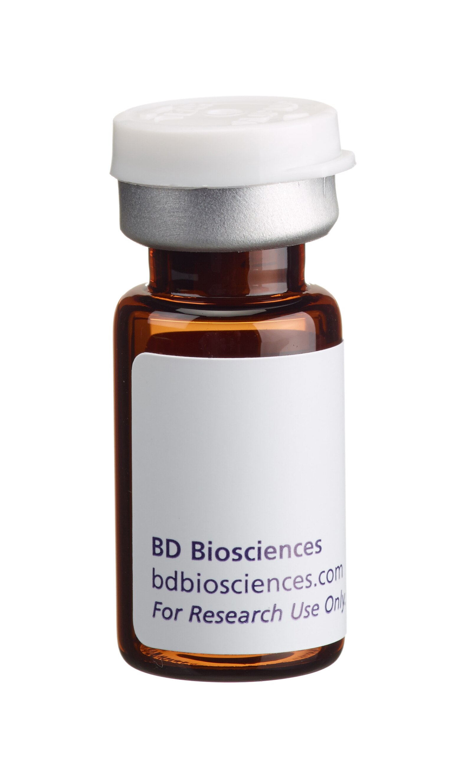
BD Pharmingen™ Ac-DEVD-CHO Caspase-3 Inhibitor
(RUO)


Spectrofluorometric analysis of Caspase-3 activity. Lysates were prepared from Daudi B cells untreated (left panel)) or treated with anti-human Fas antibody, clone DX2 (Cat. No. 555670) and Protein G (middle and right panels) for 4 hours to induce apoptosis. Cells were incubated with the Ac-DEVD-AMC, Caspase-3 substrate (left and middle panels) or the substrate and the Ac-DEVD-CHO inhibitor (right panel) and analyzed by spectrofluorometry. Left panel: Lysates from untreated cells did not emit fluorescence, indicating that the substrate was not cleaved and hence Caspase-3 activity was absent. Middle panel: Lysates from cells treated with anti-Fas mAb cleaved the substrate, indicating that presence of Caspase-3 activity. Right panel: Fluorescence was not emitted in lysates from cells treated with anti-Fas mAb when both the inhibitor and substrate were added, indicating that Caspase-3 activity was blocked. The addition of Protein G enhances the ability of clone DX2 to induce apoptosis, presumably by cross-linking Fas receptors.


BD Pharmingen™ Ac-DEVD-CHO Caspase-3 Inhibitor

Regulatory Status Legend
Any use of products other than the permitted use without the express written authorization of Becton, Dickinson and Company is strictly prohibited.
Preparation And Storage
Reconstitute the substrate before use. Reconstitute in 1 ml DMSO to yield 1 mg/ml peptide in DMSO.Store the reconstituted substrate at –20°C for up to 1-2 months and avoid repeated freeze-thaw cycles, which greatly alter product stability.
Recommended Assay Procedures
The Ac-DEVD-CHO inhibitor is designed to be used in protease assays, like those described in the literatureby Mashima et al and
Nicholson et al. When Ac-DEVD-AMC is treated with apoptotic cell lysates, AMC is released. AMC release is monitored in a spectrofluorometer at an excitation wavelength of 380 nm and an emission wavelength range of 430-460 nm. Apoptotic cell lysates yield a considerable emission as compared to non-apoptotic cell lysates or apoptotic lysates in presence of Ac-DEVD-CHO. Apoptosis can be induced by a number of mechanisms. For each protease assay, we typically use 1 ml reaction buffer [20 mM HEPES (pH 7.5), 10% glycerol, 2 mM DTT], 20 µM (final concentration) Ac-DEVD-AMC, 100 nM Ac-DEVD-CHO, and cell lysate. The amount of cell lysate required for protease assays will vary between experimental systems and should be optimized by the user.
AC-DEVD-CHO PROTEASE ASSAY
The Protease Assay protocol is used at BD Pharmingen™ to assay protease activity using the Ac-DEVD-CHO, Caspase-3 (CPP32) Inhibitor (Cat. No. 556465).
Materials Required
1. Apoptotic and non-apoptotic cells.
2. Phosphate Buffered Saline (PBS) Wash Buffer: 140 mM NaCl, 2.7 mM KCl, 10 mM KH2PO4 dissolved in distilled, autoclaved water. Adjust the pH to 7.4 using hydrochloric acid.
3. Cell Lysis Buffer: 10 mM Tris (pH 7.5), 130 mM NaCl, 1% Triton -X-100, 10 mM NaPi, 10 mM NaPPi. The Lysis Buffer is available ready-to-use (Cat. No. 554778, 50 ml).
4. Caspase-3 (CPP32), Ac-DEVD-CHO Inhibitor, (Cat. No. 556465) and substrate Ac-DEVD-AMC (Cat. No. 556449).
5. Protease Assay Buffer: 20 mM HEPES (pH 7.5), 10% glycerol, 2 mM DTT.
Procedure
Cell lysate preparation: For adherent cells, decant the media and wash rapidly with PBS. Remove excess PBS by aspiration and lyse with Cell Lysis Buffer (~2 million cells/ml). For cells growing in suspension, pellet, was with PBS and lyse with Cell Lysis Buffer (~2 million cells/ml).
Protease assays:
1. For each reaction, add 20 µM (final concentration) Ac-DEVD-AMC or 20 µM (final concentration) Ac-DEVD-AMC and 100 nM (final concentration) Ac-DEVD-CHO and cell lysate to 1 ml Protease Assay Buffer. The amount of apoptotic cell lysate required to cleave the Ac-DEVD-AMC will vary for each experimental system and should be titrated. We generally titrate between 10-100 µl of cell lysate per 1 ml Protease Assay Buffer.
Suggested controls: Reaction mixtures with non-apoptotic cells lysates. In these lysates, there may be a basal level of apoptosis. Reaction mixtures with lysis buffer only (no cells) are also suggested as negative controls.
2. Incubate the reaction mixtures for 2 hr at 37°C.
3. Measure the AMC liberated from the Ac-DEVD-AMC using a spectrofluorometer with an excitation wavelength of 380 nm and an emission wavelength range of 430-460 nm. Apoptotic cell lysates containing Ac-DEVD-AMC cleaving activity yield a considerable emission, which is blocked by addition of Ac-DEVD-CHO (see figure).
Product Notices
- Since applications vary, each investigator should titrate the reagent to obtain optimal results.
- Please refer to www.bdbiosciences.com/us/s/resources for technical protocols.
Members of the ICE/CED-3 cysteine protease family have key roles in inflammation and mammalian apoptosis. The ICE family member Caspase-3 (also known as CPP32, Yama, apopain) is activated early in apoptosis and appears to be involved in the proteolysis of several important molecules, including poly (ADP ribose) polymerase (PARP). Activated Caspase-3 cleaves PARP from its 116 kD to an 85 kD residual fragment. The cleavage site in PARP is C-terminal to Asp-216. The upstream sequence of the cleavage site, DEVD (Asp-Glu-Val-Asp), is utilized as a basis for the highly specific Caspase-3 substrate, Ac-DEVD-AMC and Caspase-3 inhibitor, Ac-DEVD-CHO.
Ac-DEVD-CHO is a synthetic tetrapeptide inhibitor for Caspase-3 (CPP32) and contains the amino acid sequence of the PARP cleavage site. The tetrapeptide inhibitor can be used to identify and quantify the Caspase-3 activity in apoptotic cells, and to study events downstream of Caspase-3 activation.
Development References (3)
-
Mashima T, Naito M, Kataoka S, Kawai H, Tsuruo T. Aspartate-based inhibitor of interleukin-1 beta-converting enzyme prevents antitumor agent-induced apoptosis in human myeloid leukemia U937 cells. Biochem Biophys Res Commun. 1995; 209(3):907-915. (Biology). View Reference
-
Nicholson DW, Ali A, Thornberry NA, et al. Identification and inhibition of the ICE/CED-3 protease necessary for mammalian apoptosis. Nature. 1995; 376(6535):17-18. (Biology). View Reference
-
Patel T, Gores GJ, Kaufmann SH. The role of proteases during apoptosis. FASEB J. 1996; 10(5):587-597. (Biology). View Reference
Please refer to Support Documents for Quality Certificates
Global - Refer to manufacturer's instructions for use and related User Manuals and Technical data sheets before using this products as described
Comparisons, where applicable, are made against older BD Technology, manual methods or are general performance claims. Comparisons are not made against non-BD technologies, unless otherwise noted.
For Research Use Only. Not for use in diagnostic or therapeutic procedures.
Report a Site Issue
This form is intended to help us improve our website experience. For other support, please visit our Contact Us page.