Skip to main content Skip to navigation
V450 Mouse Anti-Mouse NK-1.1
Alert icon
This SKU will be discontinuing Apr 2024. Suggested alternate SKU is [562921] or for additional support, contact your local applications specialist. Contact Us #
V450 Mouse Anti-Mouse NK-1.1
Flow cytometric analysis of NK1.1 on mouse splenocytes.  Splenocytes from C57BL/6 mice were stained with the BD Horizon™ V450 Mouse Anti-Mouse NK1.1 antibody in conjunction with a PE Hamster Anti-Mouse CD3e antibody.  Contour plots were derived from gated events based on light scattering characteristics for splenocytes.  Flow cytometry was performed on a BD LSR™ II flow cytometry system.
V450 Mouse Anti-Mouse NK-1.1
Flow cytometric analysis of NK1.1 on mouse splenocytes.  Splenocytes from C57BL/6 mice were stained with the BD Horizon™ V450 Mouse Anti-Mouse NK1.1 antibody in conjunction with a PE Hamster Anti-Mouse CD3e antibody.  Dot plots were derived from gated events based on light scattering characteristics for splenocytes.  Flow cytometry was performed on a BD LSR™ II flow cytometry system.
V450 Mouse Anti-Mouse NK-1.1
Flow cytometric analysis of NK1.1 on mouse splenocytes.  Splenocytes from C57BL/6 mice were stained with a BD Horizon™ V450 Mouse IgG2a, κ isotype control in conjunction with a PE Hamster Anti-Mouse CD3e antibody.  Dot plots were derived from gated events based on light scattering characteristics for splenocytes.  Flow cytometry was performed on a BD LSR™ II flow cytometry system.
Flow cytometric analysis of NK1.1 on mouse splenocytes.  Splenocytes from C57BL/6 mice were stained with the BD Horizon™ V450 Mouse Anti-Mouse NK1.1 antibody in conjunction with a PE Hamster Anti-Mouse CD3e antibody.  Contour plots were derived from gated events based on light scattering characteristics for splenocytes.  Flow cytometry was performed on a BD LSR™ II flow cytometry system.
Flow cytometric analysis of NK1.1 on mouse splenocytes.  Splenocytes from C57BL/6 mice were stained with the BD Horizon™ V450 Mouse Anti-Mouse NK1.1 antibody in conjunction with a PE Hamster Anti-Mouse CD3e antibody.  Dot plots were derived from gated events based on light scattering characteristics for splenocytes.  Flow cytometry was performed on a BD LSR™ II flow cytometry system.
Flow cytometric analysis of NK1.1 on mouse splenocytes.  Splenocytes from C57BL/6 mice were stained with a BD Horizon™ V450 Mouse IgG2a, κ isotype control in conjunction with a PE Hamster Anti-Mouse CD3e antibody.  Dot plots were derived from gated events based on light scattering characteristics for splenocytes.  Flow cytometry was performed on a BD LSR™ II flow cytometry system.
Product Details
Down Arrow Up Arrow


BD Horizon™
Klrb1b, CD161b, Nkrp1b; Klrb1c, CD161c, NK1.1, Nkrp1c
Mouse (QC Testing)
Mouse C3H x BALB/c IgG2a, κ
Mouse NK-1+ Spleen and Bone Marrow Cells
Flow cytometry (Routinely Tested)
0.2 mg/ml
AB_1727570
Aqueous buffered solution containing protein stabilizer and ≤0.09% sodium azide.
RUO


Preparation And Storage

Store undiluted at 4°C and protected from prolonged exposure to light. Do not freeze. The monoclonal antibody was purified from tissue culture supernatant or ascites by affinity chromatography. The antibody was conjugated with BD Horizon™ V450 under optimum conditions, and unreacted BD Horizon™ V450 was removed.

Product Notices

  1. Since applications vary, each investigator should titrate the reagent to obtain optimal results.
  2. An isotype control should be used at the same concentration as the antibody of interest.
  3. BD Horizon V450 has a maximum absorption of 406 nm and maximum emission of 450 nm. Before staining with this reagent, please confirm that your flow cytometer is capable of exciting the fluorochrome and discriminating the resulting fluorescence.
  4. Caution: Sodium azide yields highly toxic hydrazoic acid under acidic conditions. Dilute azide compounds in running water before discarding to avoid accumulation of potentially explosive deposits in plumbing.
  5. Pacific Blue™ is a trademark of Molecular Probes, Inc., Eugene, OR.
  6. For fluorochrome spectra and suitable instrument settings, please refer to our Multicolor Flow Cytometry web page at www.bdbiosciences.com/colors.
  7. Please refer to www.bdbiosciences.com/us/s/resources for technical protocols.
560524 Rev. 1
Antibody Details
Down Arrow Up Arrow
PK136

In the mouse, at least three members of the Klrb (Killer cell lectin-like receptor, subfamily b; formerly NKR-P1) gene family have been identified (Klrb1a/NKR-P1A, Klrb1b/NKR-P1B, and Klrb1c/NKR-P1C); but in the human gene family, a single homologue has been designated KLRB1, NKR-P1A, or CD161. The KLRB1/NKR-P1 family of proteins are type-II-transmembrane C-type lectin receptors. KLRB1C/NKR-P1C activates NK-cell cytotoxicity, while KLRB1B/NKR-P1B functions as an inhibitory receptor. KLRB1B/NKR-P1B protein has intracellular Immunoreceptor Tyrosine-based Inhibitory Motif (ITIM), while KLRB1C/NKR-P1C lacks ITIM and activates via association with Fc Receptor γ chain. Strikingly, KLRB1B/NKR-P1B and KLRB1C/NKR-P1C share 96% amino acid sequence identity in their extracellular C-type lectin domains. The PK136 antibody reacts with the NK-1.1 surface antigen (CD161c) encoded by the Klrb1c/NKR-P1C gene expressed on natural killer (NK) cells in selected strains of mice (eg, C57BL, FVB/N, NZB, but not A, AKR, BALB/c, CBA/J, C3H, C57BR, C58, DBA/1, DBA/2, NOD, SJL, 129) and the CD161b antigen encoded by the Klrb1b/NKR-P1B gene expressed only on Swiss NIH and SJL mice, but not on C57BL/6. Expression of KLRB1C/NKR-P1C protein is correlated with the ability to lyse tumor cells in vitro and to mediate rejection of bone marrow allografts. The NK-1.1 marker is useful in defining NK cells; however, the antigen is also expressed on a rare, specialized population of T lymphocytes (NK-T cells) and some cultured monocytes. Plate-bound PK136 mAb, in combination with low concentrations of IL-2, induces proliferation of a subset of NK cells.

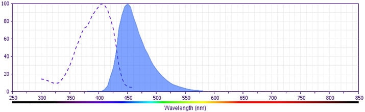
The antibody is conjugated to BD Horizon™ V450, which has been developed for use in multicolor flow cytometry experiments and is available exclusively from BD Biosciences. It is excited by the Violet laser Ex max of 406 nm and has an Em Max at 450 nm. Conjugates with BD Horizon™ V450 can be used in place of Pacific Blue™ conjugates.

560524 Rev. 1
Format Details
Down Arrow Up Arrow
V450
BD Horizon™ V450 Dye is part of the BD Horizon™ violet family of dyes. This is a small organic fluorochrome with an excitation maximum (Ex Max) at 405-nm and an emission maximum (Em Max) at 450-nm. BD Horizon™ V450, driven by BD innovation, is designed to be excited by the violet laser (405 nm) and detected using an optical filter centered near 450-nm (e.g., a 450/50-nm bandpass filter). The dye can be excited by the UV (355-nm) laser resulting in cross-laser excitation and spillover. Please ensure that your instrument’s configurations (lasers and optical filters) are appropriate for this dye.
altImg
V450
Violet 405 nm
405 nm
450 nm
560524 Rev.1
Citations & References
Down Arrow Up Arrow

Development References (15)

  1. Arase N, Arase H, Park SY, Ohno H, Ra C, Saito T. Association with FcRgamma is essential for activation signal through NKR-P1 (CD161) in natural killer (NK) cells and NK1.1+ T cells. J Exp Med. 1997; 186(12):1957-1963. (Biology). View Reference
  2. Carlyle JR, Martin A, Mehra A, Attisano L, Tsui FW, Zuniga-Pflucker JC. Mouse NKR-P1B, a novel NK1.1 antigen with inhibitory function. J Immunol. 1999; 162(10):5917-5923. (Biology: Immunoprecipitation). View Reference
  3. Giorda R, Trucco M. Mouse NKR-P1. A family of genes selectively coexpressed in adherent lymphokine-activated killer cells. J Immunol. 1991; 147(5):1701-1708. (Biology). View Reference
  4. Greimers R, Trebak M, Moutschen M, Jacobs N, Boniver J. Improved four-color flow cytometry method using fluo-3 and triple immunofluorescence for analysis of intracellular calcium ion ([Ca2+]i) fluxes among mouse lymph node B- and T-lymphocyte subsets. Cytometry. 1996; 23(3):205-217. (Biology). View Reference
  5. Karlhofer FM, Yokoyama WM. Stimulation of murine natural killer (NK) cells by a monoclonal antibody specific for the NK1.1 antigen. IL-2-activated NK cells possess additional specific stimulation pathways. J Immunol. 1991; 146(10):3662-3673. (Biology). View Reference
  6. Koo GC, Dumont FJ, Tutt M, Hackett J Jr, Kumar V. The NK-1.1(-) mouse: a model to study differentiation of murine NK cells. J Immunol. 1986; 137(12):3742-3747. (Biology). View Reference
  7. Koo GC, Peppard JR. Establishment of monoclonal anti-Nk-1.1 antibody. Hybridoma. 1984; 3(3):301-303. (Immunogen). View Reference
  8. Kung SK, Su RC, Shannon J, Miller RG. The NKR-P1B gene product is an inhibitory receptor on SJL/J NK cells. J Immunol. 1999; 162(10):5876-5887. (Biology: Blocking). View Reference
  9. Lanier LL. Natural killer cells: from no receptors to too many. Immunity. 1997; 6(4):371-378. (Biology). View Reference
  10. Reichlin A, Yokoyama WM. Natural killer cell proliferation induced by anti-NK1.1 and IL-2. Immunol Cell Biol. 1998; 76(2):143-152. (Biology: (Co)-stimulation). View Reference
  11. Sentman CL, Hackett J Jr, Moore TA, Tutt MM, Bennett M, Kumar V. Pan natural killer cell monoclonal antibodies and their relationship to the NK1.1 antigen. Hybridoma. 1989; 8(6):605-614. (Biology). View Reference
  12. Sentman CL, Kumar V, Koo G, Bennett M. Effector cell expression of NK1.1, a murine natural killer cell-specific molecule, and ability of mice to reject bone marrow allografts. J Immunol. 1989; 142(6):1847-1853. (Biology: Depletion). View Reference
  13. Vicari AP, Zlotnik A. Mouse NK1.1+ T cells: a new family of T cells. Immunol Today. 1996; 17(2):71-76. (Biology). View Reference
  14. Yokoyama WM, Seaman WE. The Ly-49 and NKR-P1 gene families encoding lectin-like receptors on natural killer cells: the NK gene complex. Annu Rev Immunol. 1993; 11:613-635. (Biology). View Reference
  15. Yu YY, Kumar V, Bennett M. Murine natural killer cells and marrow graft rejection. Annu Rev Immunol. 1992; 10:189-213. (Biology). View Reference
View All (15) View Less
560524 Rev. 1

Please refer to Support Documents for Quality Certificates


Global - Refer to manufacturer's instructions for use and related User Manuals and Technical data sheets before using this products as described


Comparisons, where applicable, are made against older BD Technology, manual methods or are general performance claims.  Comparisons are not made against non-BD technologies, unless otherwise noted.

For Research Use Only. Not for use in diagnostic or therapeutic procedures.