Skip to main content Skip to navigation
BB515 Rat Anti-Mouse CD195
Alert icon
This SKU will be discontinuing Apr 2024. For additional support, contact your local applications specialist. Contact Us #
BB515 Rat Anti-Mouse CD195
Two-color flow cytometric analysis of CD195 expression on mouse peritoneal exudate cells. Mouse peritoneal exudate cells (PEC) were isolated 3 days post-stimulation by intraperitoneal injection of a 3% thioglycollate solution. The PEC were preincubated with Purified Rat Anti-Mouse CD16/CD32 antibody (Mouse BD Fc Block™) (Cat. No. 553141/553142). The cells were then stained with APC Rat Anti-Mouse CD11b (Cat. No. 553312/561690) and either BD Horizon™ BB515 Rat IgG2c, κ Isotype Control (Cat. No. 565091; Left Panel) or BD Horizon BB515 Rat Anti-Mouse CD195 (Cat. No. 565093; Right Panel). Two-color flow cytometric contour plots showing the correlated expression of CD195 (or Ig Isotype control staining) versus CD11b were derived from gated events with the forward and side light-scatter characteristics of viable PEC. Flow cytometric analysis was performed using a BD™ LSR II Flow Cytometer System.
Product Details
Down Arrow Up Arrow


BD Horizon™
Ccr5; chemokine (C-C) receptor 5; C-C CKR-5; CC-CKR-5; CCR-5; AM4-7
Mouse (QC Testing)
Rat IgG2c, κ
Mouse CCR5 aa. 9-30
Flow cytometry (Routinely Tested)
0.2 mg/ml
AB_2739063
Aqueous buffered solution containing ≤0.09% sodium azide.
RUO


Preparation And Storage

Store undiluted at 4°C and protected from prolonged exposure to light. Do not freeze. The monoclonal antibody was purified from tissue culture supernatant or ascites by affinity chromatography. The antibody was conjugated with BD Horizon™ BB515 under optimum conditions and unconjugated antibody was removed.

Recommended Assay Procedures

BD™ CompBeads can be used as surrogates to assess fluorescence spillover (Compensation).  When fluorochrome conjugated antibodies are bound to CompBeads, they have spectral properties very similar to cells.   However, for some fluorochromes there can be small differences in spectral emissions compared to cells, resulting in spillover values that differ when compared to biological controls.  It is strongly recommended that when using a reagent for the first time, users compare the spillover on cells and CompBead to ensure that BD Comp beads are appropriate for your specific cellular application.

For optimal and reproducible results, BD Horizon Brilliant Stain Buffer should be used anytime two or more BD Horizon Brilliant dyes are used in the same experiment.  Fluorescent dye interactions may cause staining artifacts which may affect data interpretation.  The BD Horizon Brilliant Stain Buffer was designed to minimize these interactions.  More information can be found in the Technical Data Sheet of the BD Horizon Brilliant Stain Buffer (Cat. No. 563794/566349) or the BD Horizon Brilliant Stain Buffer Plus (Cat. No. 566385).

For optimal results, it is recommended to perform 2 washes after staining with antibodies. Cells may be prepared, stained with antibodies and washed twice with wash buffer per established protocols for immunofluorescence staining, prior to acquisition on a flow cytometer. Performing fewer than the recommended wash steps may lead to increased spread of the negative population.

Product Notices

  1. Since applications vary, each investigator should titrate the reagent to obtain optimal results.
  2. An isotype control should be used at the same concentration as the antibody of interest.
  3. Caution: Sodium azide yields highly toxic hydrazoic acid under acidic conditions. Dilute azide compounds in running water before discarding to avoid accumulation of potentially explosive deposits in plumbing.
  4. For fluorochrome spectra and suitable instrument settings, please refer to our Multicolor Flow Cytometry web page at www.bdbiosciences.com/colors.
  5. Please refer to http://regdocs.bd.com to access safety data sheets (SDS).
  6. BD Horizon Brilliant Stain Buffer is covered by one or more of the following US patents: 8,110,673; 8,158,444; 8,575,303; 8,354,239.
  7. Please refer to www.bdbiosciences.com/us/s/resources for technical protocols.
565093 Rev. 2
Antibody Details
Down Arrow Up Arrow
C34-3448

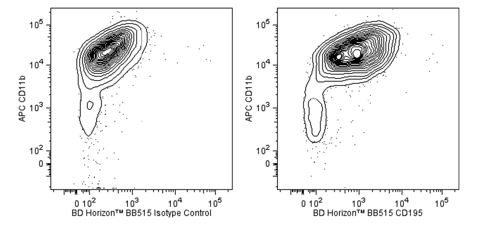
The C34-3448 monoclonal antibody specifically binds to CD195 which is also known as, C-C chemokine receptor type 5 (CCR5). CD195 is a seven transmembrane-spanning G-protein-coupled receptor that belongs to the β-chemokine receptor family. CD195 regulates lymphocyte chemotaxis activation and transendothelial migration during inflammation. It signals in response to at least three chemokines: CCL3/MIP-1α, CCL4/MIP-1β, and CCL5/RANTES. CD195 is expressed on macrophages and some T-lymphocytes.

The antibody was conjugated to BD Horizon BB515 which is part of the BD Horizon Brilliant™ Blue family of dyes. With an Ex Max near 490 nm and an Em Max near 515 nm, BD Horizon BB515 can be excited by the blue laser (488 nm) laser and detected with a 530/30 nm filter. This dye has been exclusively developed by BD Biosciences and is up to seven times brighter than FITC with less spillover into the PE channel. Due to similar excitation and emission properties, BB515, FITC, and Alexa Fluor® 488 cannot be used simultaneously. It is not recommended to use BB515 in cocktails that include Streptavidin conjugates as it may cause high background.

565093 Rev. 2
Format Details
Down Arrow Up Arrow
BB515
The BD Horizon Brilliant™ Blue 515 (BB515) dye is part of the BD Horizon Brilliant™ Blue family of dyes. This dye is a polymer fluorochrome with an excitation maximum (Ex Max) at 490-nm and an emission maximum (Em Max) of 515-nm. Driven by BD innovation, BB515 is designed to be excited by the blue laser (488-nm) and detected using an optical filter centered near 520-nm (e.g., 530/30-nm). BB515 reagents are significantly brighter than equivalent FITC or Alexa Fluor™ 488 reagents with less spillover into the PE detector. Please ensure that your instrument’s configurations (lasers and optical filters) are appropriate for this dye.
altImg
BB515
Blue 488 nm
490 nm
515 nm
565093 Rev.2
Citations & References
Down Arrow Up Arrow

Development References (10)

  1. Boring L, Gosling J, Monteclaro FS, Lusis AJ, Tsou CL, Charo IF. Molecular cloning and functional expression of murine JE (monocyte chemoattractant protein 1) and murine macrophage inflammatory protein 1alpha receptors: evidence for two closely linked C-C chemokine receptors on chromosome 9. J Biol Chem. 1996; 271(13):7551-7558. (Biology). View Reference
  2. Hayakawa Y, Smyth MJ. CD27 dissects mature NK cells into two subsets with distinct responsiveness and migratory capacity. J Immunol. 2006; 176(3):1517-1524. (Clone-specific: Flow cytometry). View Reference
  3. Kim SH, Cleary MM, Fox HS, Chantry D, Sarvetnick N. CCR4-bearing T cells participate in autoimmune diabetes. J Clin Invest. 2002; 110(11):1675-1686. (Clone-specific: Flow cytometry). View Reference
  4. McGuirk P, McCann C, Mills KH. Pathogen-specific T regulatory 1 cells induced in the respiratory tract by a bacterial molecule that stimulates interleukin 10 production by dendritic cells: a novel strategy for evasion of protective T helper type 1 responses by Bordetella pertussis. J Exp Med. 2002; 195(2):221-231. (Clone-specific: Flow cytometry). View Reference
  5. McLoughlin RM, Jenkins BJ, Grail D, et al. IL-6 trans-signaling via STAT3 directs T cell infiltration in acute inflammation. Proc Natl Acad Sci U S A. 2005; 102(27):9589-9594. (Clone-specific: Flow cytometry). View Reference
  6. Meyer A, Coyle AJ, Proudfoot AE, Wells TN, Power CA. Cloning and characterization of a novel murine macrophage inflammatory protein-1 alpha receptor. J Biol Chem. 1996; 271(24):14445-14451. (Biology). View Reference
  7. Nakae S, Iwakura Y, Suto H, Galli SJ. Phenotypic differences between Th1 and Th17 cells and negative regulation of Th1 cell differentiation by IL-17. J Leukoc Biol. 2007; 81(5):1258-1268. (Clone-specific: Flow cytometry). View Reference
  8. Napolitano M, Seamon KB, Leonard WJ. Identification of cell surface receptors for the Act-2 cytokine. J Exp Med. 1990; 172(1):285-289. (Biology). View Reference
  9. Zozulya AL, Reinke E, Baiu DC, Karman J, Sandor M, Fabry Z. Dendritic cell transmigration through brain microvessel endothelium is regulated by MIP-1alpha chemokine and matrix metalloproteinases. J Immunol. 2007; 178(1):520-529. (Clone-specific: Flow cytometry). View Reference
  10. van Deventer HW, Wu QP, Bergstralh DT, et al. C-C chemokine receptor 5 on pulmonary fibrocytes facilitates migration and promotes metastasis via matrix metalloproteinase 9. Am J Pathol. 2008; 173(1):253-264. (Clone-specific: Flow cytometry). View Reference
View All (10) View Less
565093 Rev. 2

Please refer to Support Documents for Quality Certificates


Global - Refer to manufacturer's instructions for use and related User Manuals and Technical data sheets before using this products as described


Comparisons, where applicable, are made against older BD Technology, manual methods or are general performance claims.  Comparisons are not made against non-BD technologies, unless otherwise noted.

For Research Use Only. Not for use in diagnostic or therapeutic procedures.