Skip to main content Skip to navigation
Alexa Fluor® 647 Mouse Anti-Human RANTES
Two-color flow cytometric analysis of RANTES expression in human peripheral blood lymphocytes. Human peripheral blood mononuclear cells were cultured (5 hr) with Recombinant Human IFN-γ (Cat. No. 554617/554616; 10 ng/ml) and lipopolysaccharide (Sigma, Cat. No. L-8274; 1.0 μg/ml) in the presence of BD GolgiStop™ Protein Transport Inhibitor (containing Monensin) (Cat. No. 554724). The cells were harvested, washed with BD Pharmingen™ Stain Buffer (FBS) (Cat. No. 554656), and fixed with BD Cytofix™ Fixation Buffer (Cat. No. 554655).        The cells were then permeabilized and stained in BD Perm/Wash™ Buffer (Cat. No. 554723) with PE Mouse Anti-Human CD3 antibody (Cat. No. 555333/561808/561809) and either Alexa Fluor® 647 Mouse IgG1 κ Isotype Control (Cat. No. 557732/557783; Left Panel) or Alexa Fluor® 647 Mouse Anti-Human RANTES antibody (Cat. No. 564753; Right Panel) using BD Biosciences Intracellular Cytokine Staining protocol. Two-color flow cytometric contour plots showing the correlated expression of RANTES (or Ig Isotype control staining) versus CD3 were derived from gated events with the forward and side light-scatter characteristics of intact stimulated lymphocytes. Flow cytometric analysis was performed using a BD LSRFortessa™ Cell Analyzer System.
Product Details
Down Arrow Up Arrow


BD Pharmingen™
CCL5; chemokine (C-C motif) ligand 5; EoCP; SCYA5; SIS-delta; SISd; TCP228
Human (QC Testing)
Mouse BALB/c IgG1, κ
Recombinant Human RANTES Protein
Intracellular staining (flow cytometry) (Routinely Tested)
5 µl
AB_2869611
Aqueous buffered solution containing BSA and ≤0.09% sodium azide.
RUO


Preparation And Storage

Store undiluted at 4°C and protected from prolonged exposure to light. Do not freeze. The monoclonal antibody was purified from tissue culture supernatant or ascites by affinity chromatography. The antibody was conjugated to Alexa Fluor® 647 under optimum conditions, and unreacted Alexa Fluor® 647 was removed.

Product Notices

  1. This reagent has been pre-diluted for use at the recommended Volume per Test. We typically use 1 × 10^6 cells in a 100-µl experimental sample (a test).
  2. An isotype control should be used at the same concentration as the antibody of interest.
  3. Source of all serum proteins is from USDA inspected abattoirs located in the United States.
  4. Caution: Sodium azide yields highly toxic hydrazoic acid under acidic conditions. Dilute azide compounds in running water before discarding to avoid accumulation of potentially explosive deposits in plumbing.
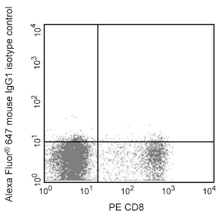
  5. Alexa Fluor® 647 fluorochrome emission is collected at the same instrument settings as for allophycocyanin (APC).
  6. The Alexa Fluor®, Pacific Blue™, and Cascade Blue® dye antibody conjugates in this product are sold under license from Molecular Probes, Inc. for research use only, excluding use in combination with microarrays, or as analyte specific reagents. The Alexa Fluor® dyes (except for Alexa Fluor® 430), Pacific Blue™ dye, and Cascade Blue® dye are covered by pending and issued patents.
  7. Alexa Fluor® is a registered trademark of Molecular Probes, Inc., Eugene, OR.
  8. For fluorochrome spectra and suitable instrument settings, please refer to our Multicolor Flow Cytometry web page at www.bdbiosciences.com/colors.
  9. Please refer to www.bdbiosciences.com/us/s/resources for technical protocols.
564753 Rev. 1
Antibody Details
Down Arrow Up Arrow
2D5

The 2D5 monoclonal antibody specifically binds to human Regulated upon Activation, Normal T cells Expressed and Secreted (RANTES). RANTES belongs to the C-C family of chemokines and is also known as CCL5. RANTES is produced by activated macrophages, CD8+ T cells, platelets, fibroblasts, epithelial cells and some tumor cells. RANTES is a chemoattractant and response modifier for a variety of cells including memory T cells, NK cells, dendritic cells, monocytes, eosinophils, basophils, and mast cells. RANTES binds to and transduces intracellular signals through several cell surface chemokine receptors including CCR1, CCR3, CCR4, and CCR5. RANTES can reportedly inhibit the interaction between certain HIV viruses and CCR5 and thus suppress viral infection in vitro.

564753 Rev. 1
Format Details
Down Arrow Up Arrow
Alexa Fluor™ 647
Alexa Fluor™ 647 Dye is part of the BD red family of dyes. This is a small organic fluorochrome with an excitation maximum (Ex Max) at 653-nm and an emission maximum (Em Max) at 669-nm. Alexa Fluor 647 is designed to be excited by the Red laser (627-640 nm) and detected using an optical filter centered near 520-nm (e.g., a 660/20 nm bandpass filter). Please ensure that your instrument’s configurations (lasers and optical filters) are appropriate for this dye.
Alexa Fluor™ 647
Red 627-640 nm
653 nm
669 nm
564753 Rev.1
Citations & References
Down Arrow Up Arrow

Development References (5)

  1. Cocchi F, DeVico AL, Garzino-Demo A, Arya SK, Gallo RC, Lusso P. Identification of RANTES, MIP-1 alpha, and MIP-1 beta as the major HIV-suppressive factors produced by CD8+ T cells. Science. 1995; 270(5243):1811-1815. (Biology). View Reference
  2. Prussin C, Metcalfe DD. Detection of intracytoplasmic cytokine using flow cytometry and directly conjugated anti-cytokine antibodies. J Immunol Methods. 1995; 188(1):117-128. (Methodology: Flow cytometry). View Reference
  3. Schall TJ, Bacon K, Toy KJ, Goeddel DV. Selective attraction of monocytes and T lymphocytes of the memory phenotype by cytokine RANTES. Nature. 1990; 347(6294):669-671. (Biology). View Reference
  4. Schall TJ, Jongstra J, Dyer BJ, et al. A human T cell-specific molecule is a member of a new gene family. J Immunol. 1988; 141(3):1018-1025. (Biology). View Reference
  5. Sticherling M, Küpper M, Koltrowitz F, et al. Detection of the chemokine RANTES in cytokine-stimulated human dermal fibroblasts.. J Invest Dermatol. 1995; 105(4):585-91. (Immunogen: Electron microscopy, ELISA, Immunocytochemistry (cytospins), Western blot). View Reference
View All (5) View Less
564753 Rev. 1

Please refer to Support Documents for Quality Certificates


Global - Refer to manufacturer's instructions for use and related User Manuals and Technical data sheets before using this products as described


Comparisons, where applicable, are made against older BD Technology, manual methods or are general performance claims.  Comparisons are not made against non-BD technologies, unless otherwise noted.

For Research Use Only. Not for use in diagnostic or therapeutic procedures.