

Preparation And Storage
Recommended Assay Procedures
For optimal and reproducible results, BD Horizon Brilliant Stain Buffer should be used anytime two or more BD Horizon Brilliant dyes are used in the same experiment. Fluorescent dye interactions may cause staining artifacts which may affect data interpretation. The BD Horizon Brilliant Stain Buffer was designed to minimize these interactions. More information can be found in the Technical Data Sheet of the BD Horizon Brilliant Stain Buffer (Cat. No. 563794/566349) or the BD Horizon Brilliant Stain Buffer Plus (Cat. No. 566385).
Product Notices
- Since applications vary, each investigator should titrate the reagent to obtain optimal results.
- An isotype control should be used at the same concentration as the antibody of interest.
- Caution: Sodium azide yields highly toxic hydrazoic acid under acidic conditions. Dilute azide compounds in running water before discarding to avoid accumulation of potentially explosive deposits in plumbing.
- Source of all serum proteins is from USDA inspected abattoirs located in the United States.
- Although hamster immunoglobulin isotypes have not been well defined, BD Biosciences Pharmingen has grouped Armenian and Syrian hamster IgG monoclonal antibodies according to their reactivity with a panel of mouse anti-hamster IgG mAbs. A table of the hamster IgG groups, Reactivity of Mouse Anti-Hamster Ig mAbs, may be viewed at http://www.bdbiosciences.com/documents/hamster_chart_11x17.pdf.
- Alexa Fluor® is a registered trademark of Molecular Probes, Inc., Eugene, OR.
- For fluorochrome spectra and suitable instrument settings, please refer to our Multicolor Flow Cytometry web page at www.bdbiosciences.com/colors.
- Cy is a trademark of GE Healthcare.
- BD Horizon Brilliant Stain Buffer is covered by one or more of the following US patents: 8,110,673; 8,158,444; 8,575,303; 8,354,239.
- BD Horizon Brilliant Violet 711 is covered by one or more of the following US patents: 8,110,673; 8,158,444; 8,227,187; 8,455,613; 8,575,303; 8,354,239.
- Please refer to www.bdbiosciences.com/us/s/resources for technical protocols.
Companion Products


The 2F1 monoclonal antibody specifically binds to KLRG1 (Killer cell Lectin-like Receptor G1), which is the mouse homolog of the rat mast cell function-associated antigen (MAFA), on all mouse strains tested (eg, AKR/J, BALB/c, C3H/HeN, C3H.SW, C57BL/6, DBA/1, SJL, 129/J). Unlike rat MAFA, which is expressed on mast cells, mouse KLRG1 is expressed on a large subset of NK cells, lymphokine-activated killer (LAK) cells, adherent LAK (A-LAK) cells, subsets of activated CD8+ T lymphocytes, and small fractions of CD4+ and CD8+ T cells, but not mast cells. The expression of KLRG1 is correlated with reduced proliferative capacity of activated T lymphocytes or reduced effector functions of activated NK cells. KLRG1 plays a role in the regulation of leucocytes of both the innate and adaptive immune system. The 2F1 mAb reportedly stains the rat basophilic leukemia cell line, RBL-2H3, which is known to express MAFA. The KLRG1 protein is an inhibitory lectin-like type II transmembrane receptor containing a cytoplasmic motif similar to ITIM (Immunoreceptor Tyrosine-based Inhibitory Motif); its ligand has not been identified KLRG1 is expressed mainly as a homodimeric molecule consisting of two N-glycosylated subunits of approximately 30-38 kDa. The level of KLRG1 expression is reduced in MHC class I-deficient mice, although direct binding of KLRG1 to MHC class I antigens could not be detected. Crosslinking of KLRG1 by 2F1 mAb reduces TCR-mediated Ca++ mobilization and cytotoxic responses (but not IFN-γ production) by CD8+ T cells and inhibits IFN-γ and TNF production and redirected lysis by NK cells.
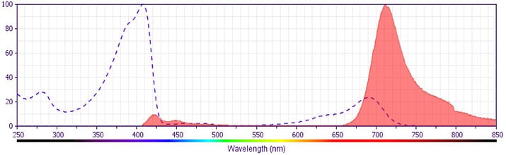
The antibody was conjugated to BD Horizon BV711 which is part of the BD Horizon Brilliant™ Violet family of dyes. This dye is a tandem fluorochrome of BD Horizon BV421 with an Ex Max of 405-nm and an acceptor dye with an Em Max at 711-nm. BD Horizon BV711 can be excited by the violet laser and detected in a filter used to detect Cy™5.5 / Alexa Fluor® 700-like dyes (eg, 712/20-nm filter). Due to the excitation and emission characteristics of the acceptor dye, there may be moderate spillover into the Alexa Fluor® 700 and PerCP-Cy5.5 detectors. However, the spillover can be corrected through compensation as with any other dye combination.
Development References (3)
-
Beyersdorf NB, Ding X, Karp K, Hanke T. Expression of inhibitory "killer cell lectin-like receptor G1" identifies unique subpopulations of effector and memory CD8 T cells. Eur J Immunol. 2001; 31(12):3443-3452. (Clone-specific: Bioassay, Blocking, Functional assay). View Reference
-
Corral L, Hanke T, Vance RE, Cado D, Raulet DH. NK cell expression of the killer cell lectin-like receptor G1 (KLRG1), the mouse homolog of MAFA, is modulated by MHC class I molecules. Eur J Immunol. 2000; 30(3):920-930. (Immunogen: Flow cytometry, Immunoprecipitation). View Reference
-
Hanke T, Corral L, Vance RE, Raulet DH. 2F1 antigen, the mouse homolog of the rat "mast cell function-associated antigen", is a lectin-like type II transmembrane receptor expressed by natural killer cells. Eur J Immunol. 1998; 28(12):4409-4417. (Clone-specific: Flow cytometry). View Reference
Please refer to Support Documents for Quality Certificates
Global - Refer to manufacturer's instructions for use and related User Manuals and Technical data sheets before using this products as described
Comparisons, where applicable, are made against older BD Technology, manual methods or are general performance claims. Comparisons are not made against non-BD technologies, unless otherwise noted.
For Research Use Only. Not for use in diagnostic or therapeutic procedures.