

Preparation And Storage
Product Notices
- Since applications vary, each investigator should titrate the reagent to obtain optimal results.
- Source of all serum proteins is from USDA inspected abattoirs located in the United States.
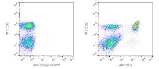
- An isotype control should be used at the same concentration as the antibody of interest.
- Please refer to www.bdbiosciences.com/us/s/resources for technical protocols.
- Alexa Fluor® is a registered trademark of Molecular Probes, Inc., Eugene, OR.
- Caution: Sodium azide yields highly toxic hydrazoic acid under acidic conditions. Dilute azide compounds in running water before discarding to avoid accumulation of potentially explosive deposits in plumbing.
- For fluorochrome spectra and suitable instrument settings, please refer to our Multicolor Flow Cytometry web page at www.bdbiosciences.com/colors.
- Cy is a trademark of Amersham Biosciences Limited.
- Brilliant Violet™ 711 is a trademark of Sirigen.
Companion Products


The DTA-1 monoclonal antibody specifically binds to GITR [Glucocorticoid-induced Tumor necrosis factor (TNF) receptor family-Related], a 66-70-kDa homodimer glycoprotein that is a member of the TNF receptor superfamily and is also known as TNFRSF18 and CD357. As its name implies, GITR expression was first detected in T lymphocytes that had been treated with dexamethasone, a glucocorticoid. In normal naive mice, GITR is expressed at moderate levels on CD25-positive/CD4-positive/CD8a-negative thymocytes and on CD25-positive/CD4-positive/CD45RB-low splenocytes. It is also expressed at low levels on splenic CD25-negative/CD4-positive/CD45RB-low T lymphocytes, B lymphocytes, macrophages, and dendritic cells. Activation of T and B lymphocytes upregulates GITR expression. GITR is a costimulatory receptor that plays an important role in Regulatory T (Treg)-cell functions, and a GITR Ligand has been detected on B lymphocytes, macrophages, and dendritic cells. mAb DTA-1 abrogates suppression by Treg cells without affecting their proliferative response, while it is co-stimulatory for T lymphocytes that are not Treg cells.
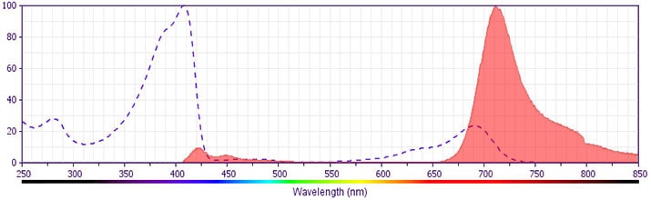
The antibody was conjugated to BD Horizon™ BV711 which is part of the BD Horizon™ Brilliant Violet™ family of dyes. This dye is a tandem fluorochrome of BD Horizon™ BV421 with an Ex Max of 405-nm and an acceptor dye with an Em Max at 711-nm. BD Horizon™ BV711 can be excited by the violet laser and detected in a filter used to detect Cy™5.5 / Alexa Fluor® 700-like dyes (eg, 712/20-nm filter). Due to the excitation and emission characteristics of the acceptor dye, there may be moderate spillover into the Alexa Fluor® 700 and PerCP-Cy™5.5 detectors. However, the spillover can be corrected through compensation as with any other dye combination.
Development References (9)
-
Beilharz MW, Sammels LM, Paun A, et al. Timed ablation of regulatory CD4-positive T cells can prevent murine AIDS progression. J Immunol. 2004; 172:4917-4925. (Biology). View Reference
-
Dittmer U, He H, Messer RJ, et al. Functional impairment of CD8-positive T cells by regulatory T cells during persistent retroviral infection. Immunity. 2004; 20:293-303. (Clone-specific: In vivo exacerbation). View Reference
-
Ji H, Liao G, Faubion WA, et al. The natural ligand for glucocorticoid-induced TNF receptor-related protein abrogates regulatory T cell suppression. J Immunol. 2004; 172:5823-5827. (Biology). View Reference
-
Kohm AP, Williams JS, Miller SD. Ligation of the glucocorticoid-induced TNF receptor enhances autoreactive CD4-positive T cell activation and experimental autoimmune encephalomyelitis. J Immunol. 2004; 172:4686-4690. (Clone-specific: Flow cytometry, In vivo exacerbation). View Reference
-
Nocentini G, Giunchi L, Ronchetti S, et al. A new member of the tumor necrosis factor/nerve growth factor receptor family inhibits T cell receptor-induced apoptosis. Proc Natl Acad Sci U S A. 1997; 94:6216-6221. (Biology). View Reference
-
Shimizu J, Moriizumi E. CD4-positive CD25-negative T cells in aged mice are hyporesponsive and exihibit suppressive activity. J Immunol. 2003; 170:1675-1682. (Clone-specific: Functional assay). View Reference
-
Shimizu J, Yamazaki S, Takahashi T, Ishida Y, Sakaguchi S. Stimulation of CD25-positive CD4-positive regulatory T cells through GITR breaks immunological self-tolerance. Nat Immunol. 2002; 3(2):135-142. (Immunogen: Cell separation, Depletion, Flow cytometry, Functional assay, Immunoprecipitation, Inhibition, In vivo exacerbation). View Reference
-
Tone M, Tone Y, Adams E, et al. Mouse glucocorticoid-induced tumor necrosis factor receptor ligand is costimulatory for T cells. Proc Natl Acad Sci U S A. 2003; 100(25):15059-15064. (Biology). View Reference
-
Uraushihara K, Kanai T, Ko K, et al. Regulation of murine inflammatory bowel disease by CD25-positive and CD25negative CD4-positive glucocorticoid-induced TNF receptor family-related gene-positive regulatory T cells. J Immunol. 2003; 171:708-716. (Clone-specific: Cell separation, Flow cytometry, In vivo exacerbation). View Reference
Please refer to Support Documents for Quality Certificates
Global - Refer to manufacturer's instructions for use and related User Manuals and Technical data sheets before using this products as described
Comparisons, where applicable, are made against older BD Technology, manual methods or are general performance claims. Comparisons are not made against non-BD technologies, unless otherwise noted.
For Research Use Only. Not for use in diagnostic or therapeutic procedures.