Preparation And Storage
Product Notices
- This reagent has been pre-diluted for use at the recommended Volume per Test. We typically use 1 × 10^6 cells in a 100-µl experimental sample (a test).
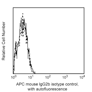
- An isotype control should be used at the same concentration as the antibody of interest.
- Source of all serum proteins is from USDA inspected abattoirs located in the United States.
- Caution: Sodium azide yields highly toxic hydrazoic acid under acidic conditions. Dilute azide compounds in running water before discarding to avoid accumulation of potentially explosive deposits in plumbing.
- Please refer to www.bdbiosciences.com/us/s/resources for technical protocols.
- This APC-conjugated reagent can be used in any flow cytometer equipped with a dye, HeNe, or red diode laser.
- For fluorochrome spectra and suitable instrument settings, please refer to our Multicolor Flow Cytometry web page at www.bdbiosciences.com/colors.
Companion Products


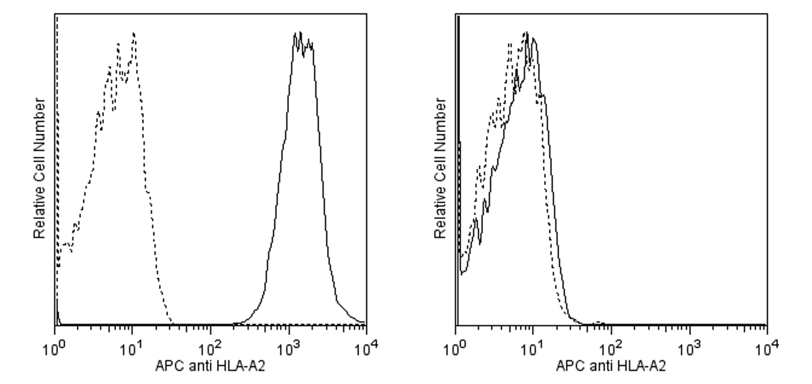
The monoclonal antibody BB7.2 specifically binds to the α subunit of the human leukocyte antigen-A2 (HLA-A2), a class I molecule of the major histocompatibility complex (MHC). The MHC gene locus encodes a group of highly polymorphic, cell-surface proteins that play a broad role in the immune response to protein antigens. MHC molecules bind and present small antigenic protein fragments to antigen-specific receptors expressed by T cells (TCR). Human (human leukocyte antigen/HLA) MHC molecules are comprised of two major classes, MHC class I and class II. Functionally, class I MHC molecules bind peptides derived from intracellular antigens (eg, viral and some bacterial antigens) which are specifically recognized by CD8+ T cells. Class II MHC molecules bind antigens derived from pathogens multiplying in intracellular vesicles and ingested extracellular bacteria, both of which are recognized by CD4+ T cells. TCR recognize processed peptides bound to the MHC as well as regions of the MHC molecule itself. CD4 and CD8 accessory molecules strengthen the formation of the TCR-MHC complex through their interaction with non-polymorphic regions of the MHC molecule.

Development References (3)
-
Bjorkman PJ, Saper MA, Samraoui B, Bennett WS, Strominger JL, Wiley DC. Structure of the human class I histocompatibility antigen, HLA-A2. Nature. 1987; 329(6139):506-512. (Biology). View Reference
-
Bjorkman PJ, Saper MA, Samraoui B, Bennett WS, Strominger JL, Wiley DC. The foreign antigen binding site and T cell recognition regions of class I histocompatibility antigens. Nature. 1987; 329(6139):512-518. (Biology). View Reference
-
Romero P, Dunbar PR, Valmori D. Ex vivo staining of metastatic lymph nodes by class I major histocompatibility complex tetramers reveals high numbers of antigen-experienced tumor-specific cytolytic T lymphocytes. J Exp Med. 1998; 188(9):1641-1650. (Biology). View Reference
Please refer to Support Documents for Quality Certificates
Global - Refer to manufacturer's instructions for use and related User Manuals and Technical data sheets before using this products as described
Comparisons, where applicable, are made against older BD Technology, manual methods or are general performance claims. Comparisons are not made against non-BD technologies, unless otherwise noted.
For Research Use Only. Not for use in diagnostic or therapeutic procedures.