
Preparation And Storage
Recommended Assay Procedures
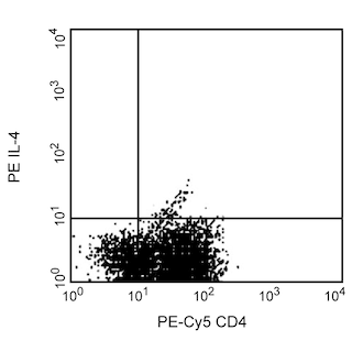
The PE-MP4-25D2 antibody is useful for immunofluorescent staining and flow cytometric analysis to identify and enumerate IL-4 producing cells within mixed cell populations (see image, left panel). For optimal immunofluorescent staining for flow cytometric analysis, this antibody should be titrated (≤ 0.20 µg mAb/million cells). For specific methodology, please visit the protocols section or chapter on Intracellular Staining in the Immune Function Handbook, both of which are posted on our web site, www.bdbiosciences.com. A useful control for demonstrating specificity of staining is the following: pre-block the fixed/permeabilized cells with unlabelled MP4-25D2 antibody (Cat. No. 554482) prior to staining. The intracellular cytokine staining technique and use of blocking controls are described in detail by C. Prussin and D. Metcalfe. A suitable rat IgG1 isotype control for assessing the level of background staining on paraformaldehyde-fixed/saponin-permeabilized mouse and human cells is PE-R3-34 immunoglobulin (Cat. No. 554685); use at comparable concentrations to antibody of interest.
OTHER APPLICATIONS
ELISA Detection: The biotinylated MP4-25D2 antibody (Cat. No. 554483) is useful as a detection antibody for a sandwich ELISA for measuring human IL-4 protein levels. For testing IL-4 in serum or plasma, the BD OptEIA™ Human IL-4 ELISA set (Cat. No. 555194) or BD OptEIA Human IL-4 ELISA Kit (Cat. No. 550614) is recommended.
Neutralization: The NA/LE™ formatted MP4-25D2 antibody (Cat. No. 554481) has been reported to be useful for neutralization of human IL-4 bioactivity.
Product Notices
- Since applications vary, each investigator should titrate the reagent to obtain optimal results.
- Please refer to www.bdbiosciences.com/us/s/resources for technical protocols.
- For fluorochrome spectra and suitable instrument settings, please refer to our Multicolor Flow Cytometry web page at www.bdbiosciences.com/colors.
- Caution: Sodium azide yields highly toxic hydrazoic acid under acidic conditions. Dilute azide compounds in running water before discarding to avoid accumulation of potentially explosive deposits in plumbing.
Companion Products

.png?imwidth=320)
The MP4-25D2 monoclonal antibody specifically binds to Interleukin-4 (IL-4). IL-4 is also known as Lymphocyte stimulatory factor 1, B cell stimulatory factor 1 (BSF-1), or B cell growth factor 1 (BCGF-1). IL-4 is produced by activated T cells, basophils, and mast cells. IL-4 is a multifunctional cytokine and growth factor that affects a variety of different target cell types. IL-4 can costimulate T cell proliferation and survival, as well as help drive Th2-like cell differentiation and effector functions. It costimulates B cell proliferation and survival, and can help B cells differentiate into IgG4- or IgE-producing cells. IL-4 can also diminish the proinflammatory functions of monocytes and macrophages. IL-4 exerts its biological effects by binding with high affinity to cell surface CD124 (IL-4Rα chain). CD124 forms signaling IL-4 receptor complexes with either CD132/common γ chain or CD213a1/IL-13Rα1 to form either Type I or Type II IL-4 Receptor complexes, respectively. The immunogen used to generate the MP4-25D2 hybridoma was purified recombinant human IL-4. This is a neutralizing antibody. The MP4-25D2 antibody has been reported to crossreact with IL-4 from rhesus monkeys. The use of the MP4-25D2 antibody for epitope mapping of human IL-4 has been described.

Development References (5)
-
Abrams JS, Roncarolo MG, Yssel H, Andersson U, Gleich GJ, Silver JE. Strategies of anti-cytokine monoclonal antibody development: immunoassay of IL-10 and IL-5 in clinical samples. Immunol Rev. 1992; 127:5-24. (Clone-specific: ELISA, Neutralization). View Reference
-
Chretien I, Van Kimmenade A, Pearce MK, Banchereau J, Abrams JS. Development of polyclonal and monoclonal antibodies for immunoassay and neutralization of human interleukin-4. J Immunol Methods. 1989; 117(1):67-71. (Clone-specific: ELISA, Neutralization). View Reference
-
Jung T, Schauer U, Rieger C, et al. Interleukin-4 and interleukin-5 are rarely co-expressed by human T cells. Eur J Immunol. 1995; 25(8):2413-2416. (Clone-specific: Flow cytometry). View Reference
-
Prussin C, Metcalfe DD. Detection of intracytoplasmic cytokine using flow cytometry and directly conjugated anti-cytokine antibodies. J Immunol Methods. 1995; 188(1):117-128. (Methodology: IC/FCM Block). View Reference
-
Ramanathan L, Ingram R, Sullivan L, et al. Immunochemical mapping of domains in human interleukin 4 recognized by neutralizing monoclonal antibodies. Biochemistry. 1993; 32(14):3549-3556. (Clone-specific: Neutralization). View Reference
Please refer to Support Documents for Quality Certificates
Global - Refer to manufacturer's instructions for use and related User Manuals and Technical data sheets before using this products as described
Comparisons, where applicable, are made against older BD Technology, manual methods or are general performance claims. Comparisons are not made against non-BD technologies, unless otherwise noted.
For Research Use Only. Not for use in diagnostic or therapeutic procedures.