Skip to main content Skip to navigation
PE Mouse Anti-Human IL-4
Expression of IL-4 by stimulated human peripheral blood mononuclear cells (PBMC). Human PBMC were stimulated with immobilized anti-human CD3 antibody (10 µg/ml for coating culture wells; Cat. No. 555329), soluble anti-human CD28 (2 µg/ml final concentration; Cat. No. 555725) recombinant human IL-2 (10 ng/ml final concentration; Cat. No. 554603) and recombinant human IL-4 (20 ng/ml final concentration; Cat. No. 554605) for 2 days. The cells were subsequently cultured in medium containing recombinant human IL-2 and recombinant human IL-4 for 3 days. Finally, the cells were harvested and stimulated for 4 hours with PMA (Sigma, Cat. #P-8139) and ionomycin (Sigma, Cat. #C-9275) in the presence of GolgiStop™ (2 µM final concentration; Cat. No. 554724). The cells were harvested, stained with FITC-anti- CD4 (Cat. No. 555346), fixed, permeabilized, and subsequently stained with 0.015 µg of PE-mouse anti-human IL-4 antibody (PE-8D4-8, Cat. No. 554516) by using Pharmingen's staining protocol (see image, left panel). To demonstrate specificity of staining, the binding of PE-8D4-8 antibody was blocked by preincubation of the antibody with recombinant human IL-4 (Cat. No. 554605; middle panel) or by preincubation of the fixed/permeabilized cells with unlabelled 8D4-8 antibody (10 µg, Cat. No. 554515; right panel) prior to staining. The quadrant markers for the bivariate dot plot were set based on the autofluorescence controls and verified using the recombinant cytokine and unlabelled antibody blocking controls.
Product Details
Down Arrow Up Arrow


BD Pharmingen™
Human (QC Testing)
Mouse IgG1, κ
Recombinant Human IL-4
Intracellular staining (flow cytometry) (Routinely Tested)
0.2 mg/ml
AB_395445
Aqueous buffered solution containing ≤0.09% sodium azide.
RUO


Preparation And Storage

Store undiluted at 4°C and protected from prolonged exposure to light. Do not freeze. The monoclonal antibody was purified from tissue culture supernatant or ascites by affinity chromatography. The antibody was conjugated with R-PE under optimum conditions, and unconjugated antibody and free PE were removed.

Recommended Assay Procedures

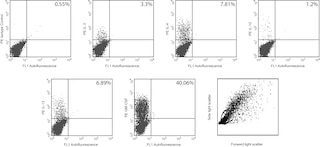
Immunofluorescent Staining and Flow Cytometric Analysis: The PE-conjugated 8D4-8 antibody can be used for multicolor immunofluorescent staining and flow cytometric analysis to identify and enumerate IL-4-producing cells within mixed cell populations (see image, left panel). For optimal immunofluorescent staining for flow cytometric analysis, titration of the antibody is strongly recommended. For specific methodology, please visit our web site, www.bdbiosciences.com, and go to the protocols section or the chapter on intracellular staining in the Immune Function Handbook. A suitable mouse IgG1 isotype control for assessing the level of background staining on paraformaldehyde - fixed/saponin -permeabilized human cells is PE-MOPC-21 (Cat. No. 554680); use at comparable concentrations to antibody of interest.

Product Notices

  1. Since applications vary, each investigator should titrate the reagent to obtain optimal results.
  2. An isotype control should be used at the same concentration as the antibody of interest.
  3. Caution: Sodium azide yields highly toxic hydrazoic acid under acidic conditions. Dilute azide compounds in running water before discarding to avoid accumulation of potentially explosive deposits in plumbing.
  4. For fluorochrome spectra and suitable instrument settings, please refer to our Multicolor Flow Cytometry web page at www.bdbiosciences.com/colors.
  5. Please refer to www.bdbiosciences.com/us/s/resources for technical protocols.
554516 Rev. 3
Antibody Details
Down Arrow Up Arrow
8D4-8

The 8D4-8 monoclonal antibody reacts with human interleukin-4 (IL-4). The immunogen used to raise the 8D4-8 hybridoma was recombinant human IL-4. The 8D4-8 antibody binds to an epitope that is different than the epitope recognized by the MP4-25D2 antibody (Cat. No. 554485).

Clone 8D4-8 displays an increased amount of non-specific binding to dead cells when compared to the clone MP4-25D2.  It is recommended to use a fixable viability dye in conjunction with this clone.

554516 Rev. 3
Format Details
Down Arrow Up Arrow
PE
R-Phycoerythrin (PE), is part of the BD family of Phycobiliprotein dyes. This fluorochrome is a multimeric fluorescent phycobiliprotein with excitation maximum (Ex Max) of 496 nm and 566 nm and an emission maximum (Em Max) at 576 nm. PE is designed to be excited by the Blue (488 nm), Green (532 nm) and Yellow-Green (561 nm) lasers and detected using an optical filter centered near 575 nm (e.g., a 575/26-nm bandpass filter). As PE is excited by multiple lasers, this can result in cross-laser excitation and fluorescence spillover on instruments with various combinations of Blue, Green, and Yellow-Green lasers. Please ensure that your instrument’s configurations (lasers and optical filters) are appropriate for this dye.
altImg
PE
Yellow-Green 488 nm, 532 nm, 561 nm
496 nm, 566 nm
576 nm
554516 Rev.3
Citations & References
Down Arrow Up Arrow

Development References (2)

  1. Bird C, Wadhwa M, Thorpe R. Development of immunoassays for human interleukin 3 and interleukin 4, some of which discriminate between different recombinant DNA-derived molecules. Cytokine. 1991; 3(6):562-567. (Clone-specific). View Reference
  2. Prussin C, Metcalfe DD. Detection of intracytoplasmic cytokine using flow cytometry and directly conjugated anti-cytokine antibodies. J Immunol Methods. 1995; 188(1):117-128. (Methodology). View Reference
554516 Rev. 3

Please refer to Support Documents for Quality Certificates


Global - Refer to manufacturer's instructions for use and related User Manuals and Technical data sheets before using this products as described


Comparisons, where applicable, are made against older BD Technology, manual methods or are general performance claims.  Comparisons are not made against non-BD technologies, unless otherwise noted.

For Research Use Only. Not for use in diagnostic or therapeutic procedures.