
Preparation And Storage
Recommended Assay Procedures
For optimal and reproducible results, BD Horizon Brilliant Stain Buffer should be used anytime two or more BD Horizon Brilliant dyes (including BD Optibuild Brilliant reagents) are used in the same experiment. Fluorescent dye interactions may cause staining artifacts which may affect data interpretation. The BD Horizon Brilliant Stain Buffer was designed to minimize these interactions. More information can be found in the Technical Data Sheet of the BD Horizon Brilliant Stain Buffer (Cat. No. 563794/566349) or the BD Horizon Brilliant Stain Buffer Plus (Cat. No. 566385).
Product Notices
- This reagent has been pre-diluted for use at the recommended Volume per Test. We typically use 1 × 10^6 cells in a 100-µl experimental sample (a test).
- Source of all serum proteins is from USDA inspected abattoirs located in the United States.
- An isotype control should be used at the same concentration as the antibody of interest.
- Caution: Sodium azide yields highly toxic hydrazoic acid under acidic conditions. Dilute azide compounds in running water before discarding to avoid accumulation of potentially explosive deposits in plumbing.
- Alexa Fluor® is a registered trademark of Molecular Probes, Inc., Eugene, OR.
- For fluorochrome spectra and suitable instrument settings, please refer to our Multicolor Flow Cytometry web page at www.bdbiosciences.com/colors.
- BD Horizon Brilliant Violet 650 is covered by one or more of the following US patents: 8,110,673; 8,158,444; 8,227,187; 8,455,613; 8,575,303; 8,354,239.
- BD Horizon Brilliant Stain Buffer is covered by one or more of the following US patents: 8,110,673; 8,158,444; 8,575,303; 8,354,239.
- Species cross-reactivity detected in product development may not have been confirmed on every format and/or application.
- Please refer to www.bdbiosciences.com/us/s/resources for technical protocols.
Companion Products


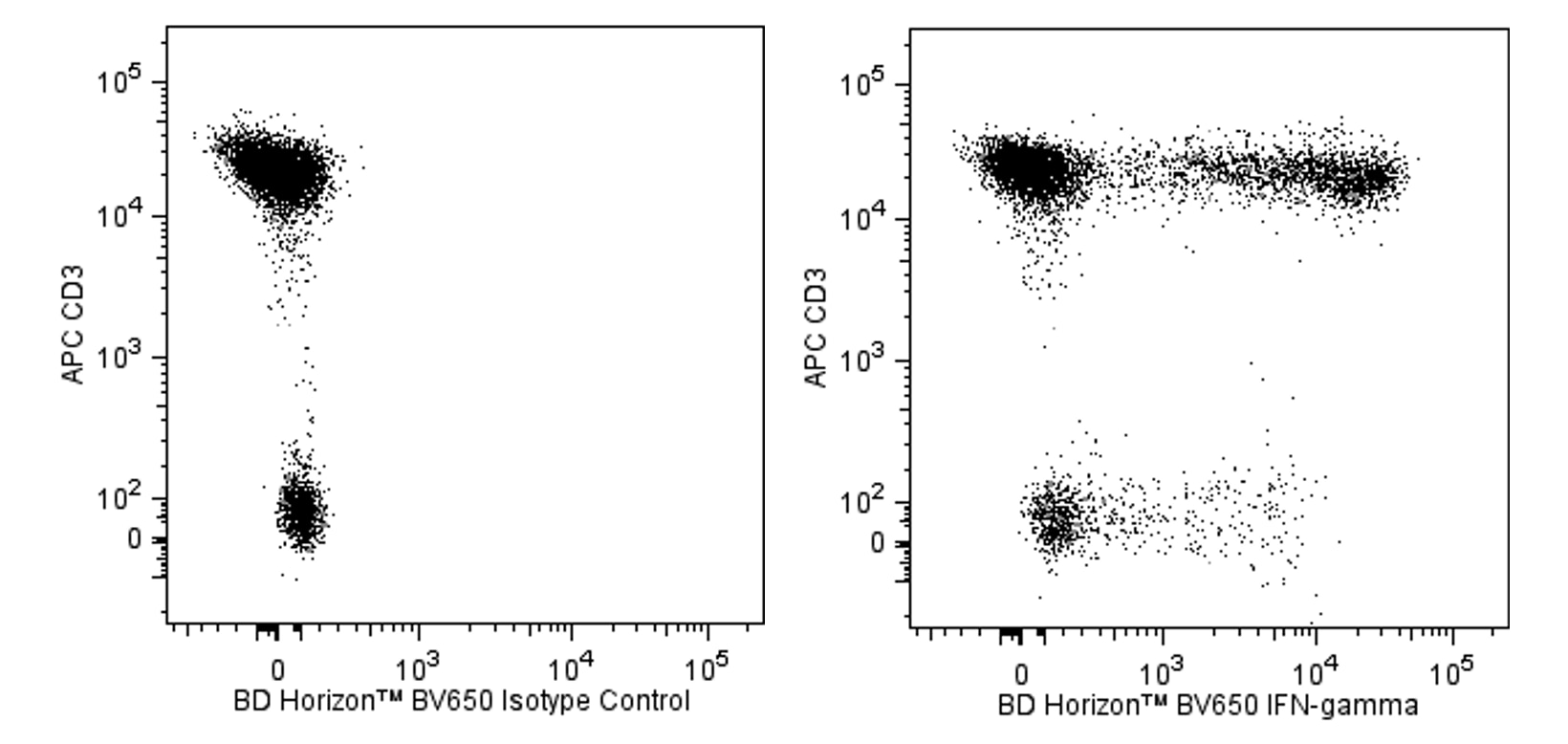
The 4S.B3 monoclonal antibody specifically binds to interferon-γ (IFN-γ). The immunogen used to generate this hybridoma was partially purified human IFN-γ obtained from supernatants of human PBMC stimulated with Staphylococcus aureus. Interferon-γ (IFN-γ) is a potent multifunctional cytokine that is produced by several activated cell types including NK, NKT, CD4+TCRαβ+, CD8+TCRαβ+, and TCRγδ+ T cells. IFN-γ exerts its biological effects through specific binding to the high-affinity IFN-γ Receptor Complex comprised of IFN-γRα (CD119) and IFN-γRβ subunits. In addition to its antiviral effects, IFN-γ upregulates a number of lymphoid cell functions including the antimicrobial and antitumor responses of macrophages, NK cells, and neutrophils. In addition, IFN-γ can exert strong regulatory influences on the proliferation, differentiation, and effector responses of B cell and T cell subsets. These influences can involve IFN-γ's capacity to boost MHC class I and II expression by antigen-presenting cells as well as to direct effects on B cells and T cells themselves. Human IFN-γ is a 14-18 kDa glycoprotein containing 143 amino acid residues.
Clone 4S.B3 also cross-reacts with a cytoplasmic component of peripheral blood CD3+ lymphocytes of baboon, and both rhesus and cynomolgus macaque monkeys following five-hour treatment with phorbol myristic acetate (PMA) and Ca++ ionophore (A23187) in the presence of monensin. The staining pattern of 4S.B3 in CD3+ cells is similar to that observed with peripheral blood T lymphocytes from normal human donors. This reagent is useful for intracellular immunofluorescent staining for flow cytometric analysis to identify and enumerate IFN-γ + cells within a mixed cell population.
The antibody was conjugated to BD Horizon™ BV650 which is part of the BD Horizon™ Brilliant Violet™ family of dyes. This dye is a tandem fluorochrome of BD Horizon™ BV421 with an Ex Max of 405-nm and an acceptor dye with an Em Max at 650-nm. BD Horizon™ BV650 can be excited by the violet laser and detected in a filter used to detect APC-like dyes (eg, 660/20-nm filter). Due to the excitation and emission characteristics of the acceptor dye, there will be spillover into the APC and Alexa Fluor® 700 detectors. However, the spillover can be corrected through compensation as with any other dye combination.

Development References (5)
-
Meager A, Parti S, Barwick S, Spragg J, O'Hagan K. Detection of hybridomas secreting monoclonal antibodies to human gamma interferon using a rapid screening technique and specificity of certain monoclonal antibodies to gamma interferon. J Interferon Res. 1984; 4(4):619-625. (Immunogen: Immunoprecipitation, Radioimmunoassay). View Reference
-
Meager A. Characterization of interferons and immunoassays. In: Clemens MJ, Morris AG, Gearing AJH, ed. Lymphokines and Interferons. A Practical Approach. Oxford: IRL Press Ltd; 1987:105-127.
-
Prussin C, Metcalfe DD. Detection of intracytoplasmic cytokine using flow cytometry and directly conjugated anti-cytokine antibodies. J Immunol Methods. 1995; 188(1):117-128. (Methodology: Flow cytometry). View Reference
-
Rotteveel FT, Kokkelink I, van Lier RA, et al. Clonal analysis of functionally distinct human CD4+ T cell subsets. J Exp Med. 1988; 168(5):1659-1673. (Clone-specific: ELISA). View Reference
-
Vogel S, Friedman R, Hogan M. Measurement of antiviral activity induced by interferons a, b, and g.. In: Coligan JE, Kruisbeek AM, Margulies DH, Shevach EM, Strober W, ed. Current Protocols in Immunology. New York: John Wiley & Sons; 2007:6.9.1-6.9.15.
Please refer to Support Documents for Quality Certificates
Global - Refer to manufacturer's instructions for use and related User Manuals and Technical data sheets before using this products as described
Comparisons, where applicable, are made against older BD Technology, manual methods or are general performance claims. Comparisons are not made against non-BD technologies, unless otherwise noted.
For Research Use Only. Not for use in diagnostic or therapeutic procedures.