

Preparation And Storage
Recommended Assay Procedures
BD® CompBeads can be used as surrogates to assess fluorescence spillover (Compensation). When fluorochrome conjugated antibodies are bound to BD® CompBeads, they have spectral properties very similar to cells. However, for some fluorochromes there can be small differences in spectral emissions compared to cells, resulting in spillover values that differ when compared to biological controls. It is strongly recommended that when using a reagent for the first time, users compare the spillover on cells and BD® CompBeads to ensure that BD® CompBeads are appropriate for your specific cellular application.
Product Notices
- Since applications vary, each investigator should titrate the reagent to obtain optimal results.
- For fluorochrome spectra and suitable instrument settings, please refer to our Multicolor Flow Cytometry web page at www.bdbiosciences.com/colors.
- Caution: Sodium azide yields highly toxic hydrazoic acid under acidic conditions. Dilute azide compounds in running water before discarding to avoid accumulation of potentially explosive deposits in plumbing.
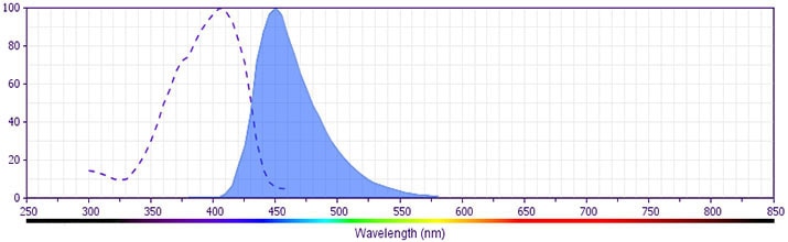
- BD Horizon V450 has a maximum absorption of 406 nm and maximum emission of 450 nm. Before staining with this reagent, please confirm that your flow cytometer is capable of exciting the fluorochrome and discriminating the resulting fluorescence.
- Please refer to www.bdbiosciences.com/us/s/resources for technical protocols.
- Please refer to http://regdocs.bd.com to access safety data sheets (SDS).
- Pacific Blue™ is a trademark of Life Technologies Corporation.
Companion Products


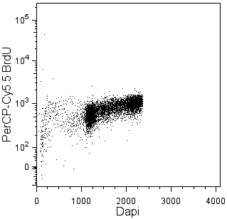
Bromodeoxyuridine (BrdU) is an analog of thymidine that can be incorporated into newly synthesized DNA by cells entering and progressing through the DNA synthesis (S) phase of the cell cycle. The amount of incorporated BrdU depends on the amount of time that the cells are exposed to BrdU (pulse time), the rate of cell division, and whether the cells are in early, mid, or late S phase. Investigators can identify cycling cells in an asynchronous cell population and determine cell cycle kinetics by detecting incorporated BrdU.
The 3D4 monoclonal antibody reacts with BrdU, but not other nucleotides, in single-stranded DNA. Random cleavage (nicking) of cellular DNA with DNase I permits the binding of the antibody to incorporated BrdU.
The antibody is conjugated to BD HorizonTM V450, which has been developed for use in multicolor flow cytometry experiments and is available exclusively from BD Biosciences. It is excited by the Violet laser Ex max of 406 nm and has an Em Max at 450 nm. Conjugates with BD HorizonTM V450 can be used in place of Pacific Blue™ conjugates.
Development References (3)
-
Dolbeare F, Gratzner H, Pallavicini MG, Gray JW. Flow cytometric measurement of total DNA content and incorporated bromodeoxyuridine. Proc Natl Acad Sci U S A. 1983; 80(18):5573-5577. (Methodology: Flow cytometry). View Reference
-
Keren DF, Hanson CA, Hurtubise PE. David F. Keren, Curtis A. Hanson, Paul E. Hurtubise., ed. Flow cytometry and clinical diagnosis. Chicago: ASCP Press; 1994:1-676.
-
Miltenburger HG, Sachse G, Schliermann M. S-phase cell detection with a monoclonal antibody. Dev Biol Stand. 1987; 66:91-99. (Clone-specific: Immunofluorescence).
Please refer to Support Documents for Quality Certificates
Global - Refer to manufacturer's instructions for use and related User Manuals and Technical data sheets before using this products as described
Comparisons, where applicable, are made against older BD Technology, manual methods or are general performance claims. Comparisons are not made against non-BD technologies, unless otherwise noted.
For Research Use Only. Not for use in diagnostic or therapeutic procedures.
Refer to manufacturer's instructions for use and related User Manuals and Technical Data Sheets before using this product as described.
Comparisons, where applicable, are made against older BD technology, manual methods or are general performance claims. Comparisons are not made against non-BD technologies, unless otherwise noted.