Old Browser
Looks like you're visiting us from {countryName}.
Would you like to stay on the current country site or be switched to your country?
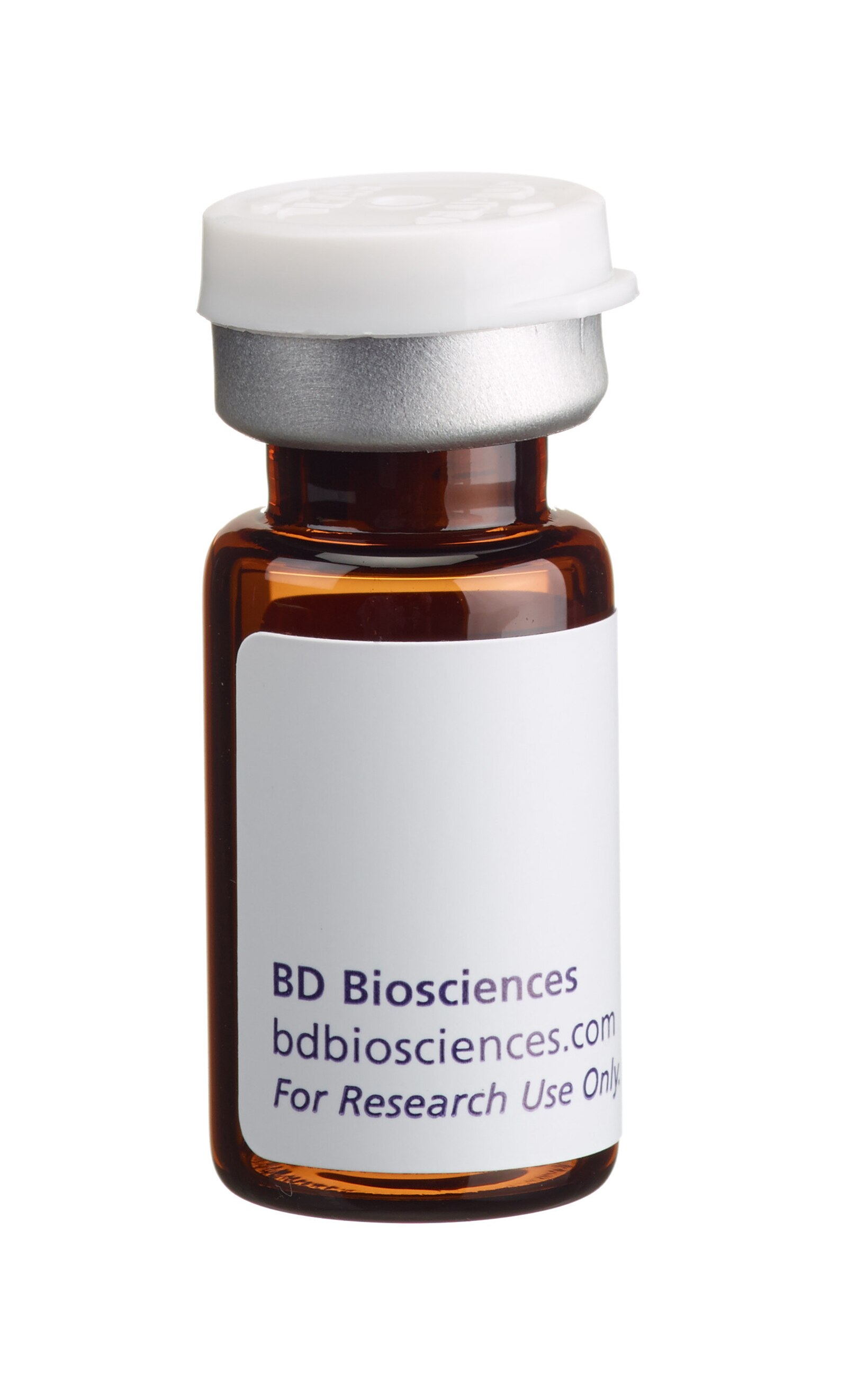
BD Pharmingen™ Hoechst 34580
(RUO)


Image analysis of live HeLa cells stained with Hoechst 34580. Live cells from the human HeLa (Cervical adenocarcinoma, ATCC CCL-2) cell line were stained with 5 μg/mL BD Pharmingen™ Hoechst 34580 (Cat. No. 565877) to segment nuclei (pseudocolored blue), 200 nM BD Pharmingen™ MitoStatus Red (Cat. No. 564697 pseudocolored red) to stain polarized mitochondria, and 10 μM Calcein AM (Cat. No. 564061, pseudocolored green) for cytoplasmic staining. The image was captured using a Molecular Devices ImageXpress® Micro XLS with a 20× objective and merged using Molecular Devices MetaXPress® software.

Flow cytometric analysis of HeLa cell DNA content using Hoechst 34580 staining. HeLa cells in log phase growth were dissociated using 0.25% Trypsin-EDTA (Life Technologies). Cells were resuspended at 1 × 10^6 cells/mL in culture medium containing 1.5 μg/mL BD Pharmingen™ Hoechst 34580 and stained (30 minutes, 37°C). Cells were pelleted, resuspended in DPBS, and analyzed by flow cytometry using a low flow rate. A 405 nm laser with a 450/50 bandpass filter was used to collect data; comparable results have also been obtained using a 355 nm laser with a 450/50 bandpass filter or a 405 nm laser with a 470/100 bandpass filter. Data was acquired using a BD LSRFortessa™ Cell Analyzer System. Histograms showing DNA levels were deconvoluted by FlowJo™ software (FlowJo, LLC) into G0/G1, S, and G2/M populations (green lines).


BD Pharmingen™ Hoechst 34580

BD Pharmingen™ Hoechst 34580

Regulatory Status Legend
Any use of products other than the permitted use without the express written authorization of Becton, Dickinson and Company is strictly prohibited.
Preparation And Storage
Recommended Assay Procedures
Preparation
Bring BD Pharmingen™ Hoechst 34580 and 1 mL distilled water to room temperature. Add 1 mL of distilled water to the vial of dye and mix well to yield a 1 mg/mL solution. Inspect the solution and repeat mixing until the stock dye has fully dissolved.
Storage
Upon arrival, store the dry dye desiccated and protected from light at ≤ -20°C until use. After reconstitution with water, store the reconstituted dye at ≤ -20°C in small aliquots. The reconstituted dye is stable for at least 6 months if stored as directed.
Procedure
Immunofluorescent Staining of Live Cells for Nuclear Visualization
1. Dilute Hoechst 34580 solution to 1-5 μg/mL in complete medium immediately prior to use.
2. Add Hoechst 34580 solution to each sample and incubate at 37°C for 30-60 minutes. The stain time required is cell type dependent.
3. Aspirate medium containing Hoechst 34580, and add BD Pharmingen™ Stain Buffer (FBS) (Cat. No. 554656) or 1× PBS. Cells may also be analyzed without washing, but this may increase background from unbound dye.
4. Proceed to immunofluorescense analysis.
5.
Immunofluorescent Staining of Fixed Cells for Nuclear Visualization
1. Fix and permeabilize cells as desired.
2. Dilute Hoechst 34580 solution to 0.5-2 µg/mL in 1× PBS immediately prior to use.
3. Add Hoechst 34580 solution to each sample at least 15 minutes before analysis.
4. Proceed to immunofluorescense analysis without washing..
5.
Staining of Live Cells for DNA Content Analysis by Flow Cytometry
1. Obtain a single cell suspension.
2. Resuspend cells at 1 × 10^6 cells/mL in complete medium containing 1-10 μg/mL Hoechst 34580. Alternatively, Hoechst 34580 may be added directly to culture without pelleting if the culture cell density does not exceed 1 × 10^6 cells/mL.
a. If staining at 1 × 10^7 cells/mL (eg, 1 × 10^6 cells in 100 μL), the recommended staining concentration is 5-10 μg/mL Hoechst 34580.
3. Incubate at 37°C for 15-60 minutes.
a. The optimal cell density, concentration of Hoechst 34580, and stain time for DNA content analysis may vary by cell type. Assay conditions should be optimized in preliminary experiments for best results.
4. Pellet cells by centrifugation and aspirate medium containing Hoechst 34580.
a. If RNA analysis (eg, RNA-seq) is to be performed after DNA content analysis is complete, wash the cells 2-3 times in serum-free buffer (eg, 1× PBS) to ensure complete removal of residual RNAse present in cell culture medium.
5. Resuspend cells in BD Pharmingen™ Stain Buffer (FBS) or 1× PBS and proceed to analysis by flow cytometry.
a. If cells are to be sorted, adding Hoechst 34580 back into the analysis buffer during acquisition at the staining concentration used may prevent dye extrusion during sorting.
b. Samples should be run at a low flow rate to achieve the best results. Increased flow rates may result in a higher % CV for each cell cycle compartment in the DNA histogram.
c. Software packages such as ModFit LT™ (Verity Software House) or FlowJo™ (FlowJo, LLC) may be useful for deconvoluting DNA content histograms into cell cycle compartments.
6.
Staining of Fixed Cells for DNA Content Analysis by Flow Cytometry
1. Obtain a single cell suspension.
2. Treat cells on ice for 30 minutes with 70-80% ice-cold ethanol.
3. Wash cells once with BD Pharmingen™ Stain Buffer (FBS).
4. Dilute Hoechst 34580 solution to 0.2-2 μg/mL in BD Pharmingen™ Stain Buffer (FBS) or 1× PBS immediately prior to use.
5. Stain cells for 15 minutes at room temperature at a cell density of 1-2 × 10^6 cells/mL. No wash is necessary prior to analysis.
a. The optimal cell density and concentration of Hoechst 34580 for DNA content analysis may vary by cell type. Assay conditions should be optimized in early experiments for best results.
6. Proceed to analysis by flow cytometry.
a. Samples should be run at a low flow rate to achieve the best results. Increased flow rates may result in a higher % CV for each cell cycle compartment in the DNA histogram.
b. Software packages such as ModFit LT™ (Verity Software House) or FlowJo™ (FlowJo, LLC) may be useful for deconvoluting DNA content histograms into cell cycle compartments.
7.
Note: Both BD Pharmingen™ Hoechst 34580 and BD Pharmingen™ Hoechst 33342 Solution (Cat. No. 561908) may be used interchangeably for most applications according to the Recommended Assay Procedure for each dye. However for DNA content analysis, Hoechst 34580 usually gives better results than Hoechst 33342 for violet laser excitation, and Hoechst 33342 usually gives better results than Hoechst 34580 for ultraviolet laser excitation.
Product Notices
- Since applications vary, each investigator should titrate the reagent to obtain optimal results.
- Please refer to www.bdbiosciences.com/us/s/resources for technical protocols.
BD Pharmingen™ Hoechst 34580 is a reagent for the fluorescent staining of DNA and nuclei in live or fixed cells. Hoechst 34580 is a bisbenzimidazole dye with high specificity for binding to double-stranded DNA. Since Hoechst 34580 is specific for DNA binding, ribonuclease treatment is not needed to avoid nonspecific RNA staining. In addition to its use in fluorescence microscopy and image analysis, Hoechst 34580 is commonly used for flow cytometric applications, such as cell cycle analysis.
Hoechst 34580 can be excited by either UV (eg, 355 or 375 nm) or violet (eg, 405 nm) light sources, and has an excitation maximum of 368 nm. It emits blue fluorescence with an emission maximum of 437 nm.
Development References (3)
-
Cherian S, Levin G, Lo WY, et al. Evaluation of an 8-color flow cytometric reference method for white blood cell differential enumeration.. Cytometry B Clin Cytom. 2010; 78(5):319-28. (Methodology). View Reference
-
Darzynkiewicz Z, Halicka HD, Zhao H. Analysis of cellular DNA content by flow and laser scanning cytometry.. Adv Exp Med Biol. 2010; 676:137-47. (Methodology). View Reference
-
Shapiro HM, Perlmutter NG. Violet laser diodes as light sources for cytometry.. Cytometry. 2001; 44(2):133-6. (Methodology). View Reference
Please refer to Support Documents for Quality Certificates
Global - Refer to manufacturer's instructions for use and related User Manuals and Technical data sheets before using this products as described
Comparisons, where applicable, are made against older BD Technology, manual methods or are general performance claims. Comparisons are not made against non-BD technologies, unless otherwise noted.
For Research Use Only. Not for use in diagnostic or therapeutic procedures.
Refer to manufacturer's instructions for use and related User Manuals and Technical Data Sheets before using this product as described.
Comparisons, where applicable, are made against older BD technology, manual methods or are general performance claims. Comparisons are not made against non-BD technologies, unless otherwise noted.
Report a Site Issue
This form is intended to help us improve our website experience. For other support, please visit our Contact Us page.